
In this episode we go through seven cases that display the breadth of presentations of limb or life threatening causes of low back pain emergencies with my huge mentors, Dr. Walter Himmel and Dr. Brian Steinhart. We cover everything from spinal epidural abscess to cauda equina syndrome to retroperitoneal bleeds, elucidating the key historical, physical exam, lab, imaging and treatment pearls for all of these diagnoses. We then go on to review the best management for the most common cause of back pain presentation, lumbosacral strain and debate the various medication options.
Dr. Steinhart & Dr. Himmel answer such questions as: Which patients with low back pain require an MRI in the ED? How useful is an ESR or CRP in diagnosing Spinal Epidural Abscess and Cord Compression secondary to spinal metastases? What is the role of dexamethasone in treating spinal cord compression? What are the physical examination maneuvers and bedside tests that are the most useful for diagnosing cauda equina syndrome and spinal cord compression?
Written summary and blog post by Claire Heslop, Edited by Anton Helman September 2012
Cite this podcast as: Himmel, W, Steinhart, B, Helman, A. Low Back Pain Emergencies. Emergency Medicine Cases. September, 2012. https://emergencymedicinecases.com/episode-26-low-back-pain-emergencies/. Accessed [date].
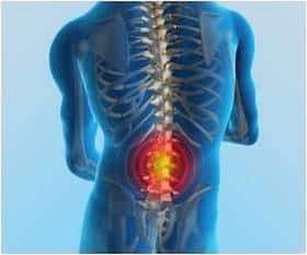
APPROACH TO LOW BACK PAIN EMERGENCIES
Main categories of patients with acute back pain:
- nonspecific lumbosacral pain/strain
- radicular pain or sciatica
- emergent pathologies.
The 5 emergent pathologies are:
- infection such as osteomyelitis, or spinal epidural abscess,
- fracture (trauma or pathologic),
- disk herniation & cord compression,
- cancer in spine causing cord compression,
- vascular – leaking/ruptured AAA, retroperitoneal bleed, and spinal epidural hematoma.
Red flags for Low Back Pain Emergencies
- Age 60,
- Symptoms or history of cancer,
- Immunodeficiency (including diabetes, IVDU), previous spinal interventions, or recent infections,
- Pain not resovled by analgesia,
- History of trauma or coagulopathy,
- Cauda equina/cord compression symptoms (bowel, bladder or erectile dysfunction, saddle paresthesia, progressive bilateral leg weakness)
Pearls: *Constant, unrelenting, severe pain, especially if it is worse lying down is a red flag for infection or cancer.* Discogenic pain is worse with flexion, and pain from spondylolysis is worse with extension
![]() Update 2018: While positive responses to “red flag’ questions for low back pain (e.g. bowel or bladder incontinence, history of cancer, trauma, fever, IV drug use etc) prompt further investigation, negative responses are not sufficient to rule out serious pathology. Though conclusions were based on a large retrospective review, the authors present data recommending caution when using “red flag” questions as screening tools. Abstract
Update 2018: While positive responses to “red flag’ questions for low back pain (e.g. bowel or bladder incontinence, history of cancer, trauma, fever, IV drug use etc) prompt further investigation, negative responses are not sufficient to rule out serious pathology. Though conclusions were based on a large retrospective review, the authors present data recommending caution when using “red flag” questions as screening tools. Abstract
![]() Update 2021: Retrospective tertiary center review (U.K.) showed that bilateral lower extremity pain, dermatomal distribution sensory loss, and loss of bilateral ankle/knee reflexes showed correlation with radiographic diagnosis of cauda equina compression (CEC); while digital rectal examination (DRE) did not demonstrate any benefit. However, individual symptoms showed poor performance in ruling in (or out) CEC. Abstract
Update 2021: Retrospective tertiary center review (U.K.) showed that bilateral lower extremity pain, dermatomal distribution sensory loss, and loss of bilateral ankle/knee reflexes showed correlation with radiographic diagnosis of cauda equina compression (CEC); while digital rectal examination (DRE) did not demonstrate any benefit. However, individual symptoms showed poor performance in ruling in (or out) CEC. Abstract
A challenge in the ED?
- Upwards of 90% of low back pain presentations in the ED are due to benign causes. However there are several important life/limb- threatening diagnoses we must consider in the low back pain patient, and most of these diagnoses are easy to miss. Furthermore, lumbosacral sprain is often associated with significant morbidity, and ED docs should provide specific education and evidence based treatments.
Physical exam for Low Back Pain Emergencies
Physical Exam Maneuvers:
- Percuss the spinous processes for tenderness, a red flag for infection and fracture,
- Test for saddle anesthesia (sensation changes may be subtle and subjective),
- DRE looking for tone/sensation
- Look for fever, or signs of infection,
- Check carefully for bilateral, or multi-level neurologic findings in lower extremities, and assess for gait disturbances.
Straight leg raise (SLR):
- Non-specific test, only positive pain is produced distal to the knee between 30–70°.
- Pain with contralateral SLR is more specific for siatica.
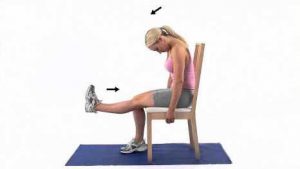
Slump test: Helps discriminate radicular pain from hamstring pain. With thoracic and cervical flexion, and knee in extension, dorsiflex the foot and flex the neck to determine if pain is produced, with release of cervical flexion to see if symptoms improve (image below).
Abdomen exam and ED ultrasound: look for AAA and bladder distention post-void.
Cognitive Forcing Strategy to Remember Serious Back Pain Pathologies:
Considering renal colic?…think about AAA!
Considering pyelonephritis?….think about spinal infection!
SPINAL EPIDURAL ABSCESS
Suspect epidural abscess in a patient with:
- back pain or neurologic deficits and fever,or
- back pain in an immune-compromised patient, or
- patient with a recent spinal procedure and either of the above
Risk Factors: Diabetes, IVDU, indwelling catheters, spinal interventions, infections elsewhere (especially skin), immune suppression (i.e. HIV), and “repeat ED visits.”
The classic triad of fever, back pain, and neurologic deficit is present in only 15% of patients, depending on stage of disease. Spinal epidural abscess is often missed on first ED visit. Fever is present in only 50% of patients, and neuro deficits start very subtly.
CRP and ESR may help, depending on the clinical suspicion for epidural abscess. If suspicion is low after the history and physical, low ESR and CRP levels support not doing an MRI, and discharging the patient home with close follow up. If there is a high index of suspicion, an MRI is indicated regardless of CRP and ESR.
Remember the normal ESR cutoff is: (age+10)/2.
In one study of epidural abscesses, 98% had ESR >20, and most were much higher (>60).
Is there any role for CT scan? CT cannot rule out epidural abscess because it does not show the epidural space, spinal cord, or spinal nerves adequately. CT can lead to the diagnosis of osteomyelitis and premature closure, missing coexistent abscess (and the urgent indication for surgery).
Remember if the suspicion is epidural abscess, the entire spine must be imaged by MRI. Spinal cord obstruction and paralysis can happen very quickly from epidural abscess, so there needs to be definitive imaging and surgical decompression as quickly as possible.
Start antibiotics while awaiting definitive diagnosis: include appropriate coverage for MSSA and MRSA, and cover gram negatives.
CAUDA EQUINA SYNDROME
Definition of Cauda Equina Syndrome:
- urinary retention or rectal dysfunction or sexual dysfunction (or all of the above)
PLUS
- saddle or anal anesthesia and/or hypoesthesia (1).
- Urinary retention is non-specific for cauda equina syndrome, but sensitive.
- A post void residual >100mL should raise the suspicion for cauda equina syndrome
When are steroids indicated?
- Evidence supports dexamethasone for metastasis to spine causing cauda equina.
- There is no indication for IV steroids for patients with cord compression by other causes
SPINAL METASTASIS – A LOW BACK PAIN EMERGENCY
- Known cancer + new back pain = spinal metastases until proven otherwise!
- Time is Limbs: Spinal metastases are one of the most common causes of cord compression. Pre-treatment neuro status predicts outcome for this emergency.
- Workup:
- X-ray to look for compression #, soft tissue changes, blastic/lytic lesions, pedicle erosion (see image below)
- Consider testing ESR and CRP, and calcium profile if signs are consistent with hypercalcemia (e.g. polyurea)
- Give dexamethasone as soon as mets are suspected (at least 10mg IV) if the patient has neurologic symptoms. Consider bisphosphonate* and calcitonin if patient is hypercalcemic, or if you suspect compression # or bony metastasis.
- Get an urgent MRI if there are symptoms of cord compression. If there are hard neurologic findings, MRI is needed within 24 hours. If the x-ray findings are consistent with mets, but there are no neuro findings, an MRI should be done within 7 days.
*Bisphosphonates may decrease bone resorption in patients with metastatic disease to the bone, and relieve pain better than placebo.
VASCULAR EMERGENCIES ARE LOW BACK PAIN EMERGENCIES
Spinal Epidural Hematoma
- Spinal epidural hematoma may present after spinal procedures (epidural anesthesia), but can be spontaneous, especially in anti-coagulated patients.
- Neurologic findings depend on the extent of spinal cord compression— from isolated pain to flaccid paralysis.
- Suspect this emergency in patients with a history of trauma and neurologic findings who are coagulopathic.
Abdominal Aortic Aneurysm
- Typical manifestations of rupture of a AAA are abdominal or back pain, with a pulsatile mass in a patient with a history of HTN.
- However, symptoms may range from dizziness, syncope, groin pain, or flank mass to presentation with paralysis. Look for livedo reticularis (atheroemboli to feet) and signs of poor circulation in the lower extremities.
- Transient hypotension or syncope after onset of pain is an important clue for bleeding from a ruptured AAA. Patients may present in shock, and quickly decline.
- Do a point of care ultrasound to rule out AAA in patients with low back pain and hypotension.
Retroperitoneal Bleed
- Patients with coagulopathies, as well as patients with retroperitoneal masses or tumors, or abdominal/pelvic trauma are at risk.
- Blood may dissect anteriorly, causing abdominal pain, or may cause pain to the hip, groin, or anterior thigh.
- On the physical exam, look for psoas sign caused by retroperitoneal irritation, femoral neuropathy and hip pain, as well as Cullen’s/Turner’s signs, or bruising or swelling in the groin caused by extension of bleeding into the skin.
LUMBOSACRAL SPRAIN & MECHANICAL LOW BACK PAIN
Lumbosacral sprain is a diagnosis of exclusion:
- Lumbosacral sprain or mechanical back pain is a diagnosis of exclusion, made only after carefully ruling out serious causes of low back pain.
Management of lumbosacral sprain:
- Education – tell the patient that “this is a mechanical problem requiring a mechanical solution – pain medications alone will not fix the problem”. Patients need to play an active role in their recovery, and studies have shown that prolonged bed rest will likely worsen the problem.
- Reassurance – 90% get better with time (over weeks)
- Symptom Management – evidence from the Cochrane collaboration supports heat, NSAIDs, acetaminophen, massage and physical therapy. Muscle relaxants may be as effective as NSAIDs, but they have significant side effects, especially in combination with opioids.
![]() Update 2017: New ACP guidelines based on a systematic review from the Annals of Internal Medicine:
Update 2017: New ACP guidelines based on a systematic review from the Annals of Internal Medicine:
1. Acute low back pain: recommends superficial heat and if pharmacological treatment is desired – NSAIDs or skeletal muscle relaxants. There was no added benefit of tramadol or opioids combined with NSAIDs for acute pain. Acetaminophen was found to be ineffective. 2. Chronic low back pain: recommends exercise, acupuncture, and mindfulness-based stress reduction while first line for pharmacological treatment continued to be NSAIDs and second line either tramadol or duloxetine. Abstract
![]() Update 2021: Prospective, randomized study on 112 admitted ED patients with low back pain (LBP) of unclear chronicity, who received intravenous dexketoprofen (NSAID). Found that intradermal sterile water injection (ISWI) was more effective in relieving LBP pain than systemic intravenous NSAID alone. Abstract
Update 2021: Prospective, randomized study on 112 admitted ED patients with low back pain (LBP) of unclear chronicity, who received intravenous dexketoprofen (NSAID). Found that intradermal sterile water injection (ISWI) was more effective in relieving LBP pain than systemic intravenous NSAID alone. Abstract
Dr. Steinhart, Dr. Himmel and Dr. Helman have no conflicts of interest to declare
For More on Back Pain on EM Cases:
Best Case Ever 11: Cauda Equina Syndrome
Key References
Armingeat,T et al. Osteoporos Int. 2006;17:1659–65. www.ncbi.nlm.nih.gov/pubmed/16896508
Davis, D et al. J Neurosurg Spine. 2011;14:765–70 www.ncbi.nlm.nih.gov/pubmed/21417700
http://back.cochrane.org/our-reviews
Lavy, C et al. BMJ. 2009;338:b926. www.ncbi.nlm.nih.gov/pubmed/19336488
Now test your knowledge with a quiz.

[…] Canadian FOAMed Reference: Emergency Medicine Cases: Low Back Pain Emergencies […]
Absolutely great…brilliant…i envy ur mentors