


How do you feel when you pick up an ED chart with a chief complaint of “Vertigo”? Do you feel confident in your approach and assessment? Do you know what to look for on your vertigo-specific tests? At the end of the day, regardless of how you feel about vertigo, these patients are coming for you on shift, and it’s worth equipping yourself with the tools to confidently manage them. Dizziness and vertigo are common ED presentations, yet there are so many myths and misperceptions that can lead us astray and create confusion when assessing these patients. So how can we take an organized and simple approach to a complex presentation like vertigo? In this Vertigo Part 1 episode, Dr. Katie Lin is joined by guest experts Dr. Peter Johns and Dr. Elisha Targonsky to tackle this tricky topic. Tune in to learn more about: common pitfalls and pearls for determining what kind of vertigo syndrome you’re dealing with and how they differ in their risk stratification, why symptom descriptors alone are not enough and how to better assess a timing and triggers history, how to classify vertigo syndromes to organize your differential diagnosis and assessment approach, what to do for episodic vertigo physical exam maneuvers, and how to treat and safely disposition your episodic vertigo patients. All this and more… (and don’t forget to tune in for our upcoming Vertigo Part 2 episode, where we’ll cover all things continuous vertigo).
Podcast: Play in new window | Download (Duration: 1:37:12 — 89.1MB)
Subscribe: Apple Podcasts
Podcast production, sound design & editing by Katie Lin & Anton Helman
Written Summary and blog post by Katie Lin, edited by Anton Helman October, 2026
Cite this podcast as: Lin, K., Johns, P., Targonsky E, Helman, A. Vertigo Part 1: Approach to Episodic Vertigo. Emergency Medicine Cases. October, 2026. https://emergencymedicinecases.com/episodic-vertigo. Accessed October 20, 2026
Dizziness and vertigo account for approximately 2–5% of emergency department (ED) visits. The assessment can be challenging because patients use imprecise and shifting descriptions, and red flags may be subtle. The vast majority of vertigo we see in the ED is due to non-life-threatening peripheral causes (eg. Benign Paroxysmal Positional Vertigo, BPPV, or vestibular neuritis), but about 3-6% of cases1 can have a serious central cause, of which posterior circulation ischemic stroke is the most dangerous diagnosis to miss.
Defining Dizziness and Vertigo
“Dizziness” is a highly non-specific term that can mean many different things to different patients. While we are often taught to ask “what do you mean by dizzy?”, literature shows that ~50% of ED patients with dizziness will change their description of the dizziness within the same visit!
For consistency of understanding, there are official definitions of dizziness, vertigo, disequilibrium, and ataxia according to the International Classification of Vestibular Disorders (ICVD):
- DIZZINESS: the sensation of disturbed or impaired spatial orientation (However, patients often also use this non-specific terminology when describing lightheadedness, feeling faint, “brain fog”, or even nausea).
- VERTIGO: the sensation of self-motion (head or body) when no self-motion is occurring or the sensation of distorted self-motion during an otherwise normal head movement.
- DYSEQUILIBRIUM (UNSTEADINESS): the feeling of being unstable when seated, standing, or walking.
- ATAXIA: a lack of voluntary coordination of muscle movements (can be truncal or limb-involving).
Clinical Pitfall: Asking “what do you mean by dizzy?” is only a starting point to understanding what symptoms the patient is experiencing, but it is not enough without additional assessment clues. Don’t overly rely on the patient’s description of their dizziness alone to help you risk stratify.
A More Helpful Approach to Dizziness Characterization
Two broad categories of dizziness that are helpful to characterize:
- Sensation of movement: spinning or imbalance sensation – potentially more suggestive of vestibular pathology
- Sensation of loss of consciousness: lightheaded, faint, or tunnel-vision – potentially more suggestive of presyncope
Instead of relying on descriptor labels of the dizziness alone, also ask about timing, triggers, episode duration, and associated features on history, similar to the approach used for other common ED complaints to help you narrow down your approach and differential diagnosis:
- Timing: When did symptoms begin? Are they still present now? How long does the intense episode last? Have there been prior episodes?
- Triggers: What was the patient doing immediately before onset? Do symptoms begin with rolling over in bed, lying down, bending forward, sitting up, standing, or another position change?
- Episode Duration/Between-Episode Symptoms: Are the symptoms persistent over time, even when the patient is holding still/not moving? Or does the patient return to normal/baseline, remain vaguely unwell, nauseated, or unsteady between episodes? Distinguish the duration of the spinning itself from the longer period of residual nausea or malaise.
- Associated features: Ask about vomiting, gait difficulty, hearing change, headache, neck pain, focal neurologic symptoms, and non-neurologic symptoms that may suggest a systemic cause.
A patient who says they have been “dizzy all day” may actually be experiencing brief, severe attacks with milder nonspecific symptoms between. Ask what they were doing at the exact moment symptoms started, whether the intensity remains constant or waxes and wanes, and whether they are currently symptomatic at rest while being assessed in the ED (and does it feel the same or different from the more severe episodic periods). For example, repeated 10–30 second attacks when rolling over or sitting up, with improvement when still, are compatible with triggered episodic vertigo, even if there is still nonspecific nausea or malaise in between severe episodes.
Clinical pearl: movement-provoked worsening is NOT proof of a peripheral disorder. Central causes, including stroke, can also worsen when the patient moves. The key is whether the vestibular symptoms are TRIGGERED by the head movement in the first place (not whether they are worsened by it).
Peripheral vs Central Causes of Vertigo & Dizziness
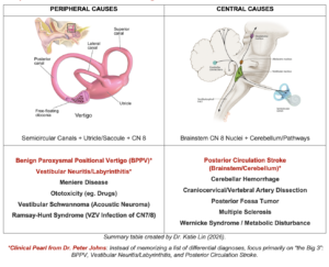
ED BEDSIDE APPROACH TO EPISODIC VERTIGO ASSESSMENT
There are many different vertigo approach algorithms that exist (eg. TiTrATE Timing and Triggers, ATTEST, STANDING, etc) with various pros and cons to each. Where all vertigo algorithms agree is that screening for red flag clues for central causes and distinguishing episodic from continuous vertigo and dizziness are key components that guide overall assessment.
Dr. Peter Johns stresses the importance of EM clinicians knowing how to recognize the “Big 3 Vertigo Diagnoses”: Benign Paroxysmal Positional Vertigo (BPPV, episodic vertigo), Vestibular Neuritis (VN, continuous vertigo), and Posterior Circulation TIA/Stroke (episodic or continuous vertigo).
This Vertigo Part 1 episode will walk through a simplified 4-step approach to episodic vertigo, while the upcoming Vertigo Part 2 episode will walk through applying the same 4-step approach to continuous vertigo.
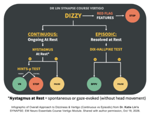
STEP 1: Screen for Central & Systemic Red Flags of Vertigo
A structured ED screening neuro exam is often more important than any specific vestibular maneuvers. Pay attention to abnormal vital signs and focal neurologic deficits. Truly isolated vertigo without any other red flag findings has a <0.7% probability of being caused by posterior circulation ischemic stroke or TIA; a solid history and screening neuro exam can do most of the heavy lifting for vertigo risk stratification, but only if we’re careful not to miss subtle clues.
Red Flags for a Central Cause
- New focal (esp unilateral) weakness, sensory change, speech disturbance, and Dangerous D’s of brainstem findings (diplopia, dysphagia, dysphonia, dysmetria, or dystaxia/marked incoordination).
- New inability to sit upright, stand, or walk without assistance, particularly when this is a change from baseline.
- Spontaneous vertical nystagmus at rest or direction-changing nystagmus at rest (ie. with the head holding still).
- New severe headache, unilateral neck pain, or significant recent head trauma, which may raise concern for posterior circulation stroke or vertebral artery dissection.
- New hearing loss or visual field deficit. Patients may not recognize or volunteer these symptoms, so ask and assess deliberately.
Gait testing is a key part of risk stratification. Ask the patient to sit up independently, then stand and walk without support if safe. Observe whether they can walk in a straight line and turn. Peripheral vestibular disorders can cause imbalance, so interpret gait in context (eg. young patient with good baseline balance vs frail elderly patient with baseline balance deficits). However, inability to stand or walk unaided and/or significant departure from baseline independent gait is a major red flag for a potential central cause.
Clinical pearl: Don’t forget to consider systemic causes and associated red flag symptoms such as: chest pain, palpitations, melena stools, febrile illness, vital sign derangements, or other evidence of systemic illness. Dr. Peter Johns notes that “dizziness + persistent hiccups” are a red flag for central causes as well.
STEP 2: Classify the Vertigo Syndrome Using a Timing & Triggers Approach
The GRACE-3 guideline framework describes three clinical vestibular syndromes:
- Acute vestibular syndrome (AVS): continuous dizziness/vertigo, typically with spontaneous nystagmus at rest; patients may have vomiting and gait disturbance. This is the setting in which HINTS/HINTS Plus may be useful when performed by a clinician trained in its use to help distinguish peripheral causes (vestibular neuritis) from central causes (posterior circulation stroke).
- Spontaneous episodic vestibular syndrome (S-EVS): intermittent episodes lasting minutes to hours without a clear trigger. Important considerations include vestibular migraine, posterior circulation TIA, and Ménière’s disease.
- Triggered episodic vestibular syndrome (T-EVS): brief episodes provoked by a position change trigger. BPPV is the key diagnosis to identify.
Clinical pearl: Rather than memorizing a series of differential diagnoses and syndromes, you can also take Dr. Peter Johns’ “Big 3” approach and learn to recognize three specific high-value diagnoses: BPPV, vestibular neuritis, and posterior circulation stroke.
STEP 3: Choose the Correct Bedside Vertigo Test for Episodic Vertigo
Dix-Hallpike for EPISODIC Vertigo
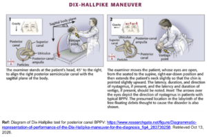
In a patient with episodic, position-triggered symptoms and no spontaneous nystagmus at rest, perform positional testing to provoke a positive BPPV finding, starting with Dix-Hallpike (since posterior canal BPPV is the most common type of BPPV).
Practical technique and interpretation:
- Begin with the patient seated. Turn the head approximately 45 degrees toward the side being tested, then lower the patient briskly so the head extends about 20–30 degrees below horizontal while remaining supported.
- Ask the patient to keep their eyes open and avoid fixating on the examiner so the eye movements can be observed. Allow time for a brief latency before calling it a “negative” test (minimum of 30-60 seconds before moving on).
- A classic positive test for posterior canal BPPV produces a transient, crescendo-decrescendo pattern of upbeat and torsional nystagmus, with the torsional component toward the downward/affected ear. It commonly settles within seconds, although it can last longer (up to 1-2 minutes).
- Return the patient to sitting position and test the other side if needed. A test without nystagmus is negative; nystagmus that does not follow the expected pattern is atypical and requires reconsideration. A test that is positive for classic findings of posterior canal BPPV should be immediately followed by an otolith repositioning maneuver (such as Epley maneuver)
If Dix-Hallpike is negative of indeterminate, but the history still suggests BPPV, consider a follow-up supine roll test for horizontal canal BPPV, looking for provoked horizontal nystagmus. The corresponding repositioning maneuver (Gufoni) is more specialized; consider using appropriate teaching resources or referral to a vestibular physiotherapist.
Dix-Hallpike Video: Dr. Peter Johns’ Youtube video on how to perform a proper Dix-Hallpike test and what a true positive test looks like can be accessed here: https://www.youtube.com/watch?v=88dPK4_8kj8
Clinical pearl: Before performing any vertigo assessment testing (including Dix-Hallpike), explain the maneuver beforehand, obtain the patient’s cooperation, and warn that it may briefly provoke intense vertigo but that the vertigo will be short-lived and the diagnosis can be made at the bedside. Consider symptom control beforehand if severe nausea is likely to prevent completion.
Do Not Misuse HINTS Plus for Episodic Vertigo
HINTS/HINTS Plus is NOT a general dizziness screen and should NOT be applied indiscriminately to all patients with vertigo. It is NOT intended for episodic vertigo without nystagmus at rest. Using HINTS in a typical BPPV presentation can produce misleading results and lead to over-investigation/over-imaging. We’ll review more about appropriate use of HINTS/HINTS Plus in Vertigo Pt 2, where we cover a simple approach to continuous vertigo.
Clinical pearl: In general, you should NOT find yourself performing both a positional maneuver like Dix-Hallpike and HINTS/HINTS Plus on the same patient.
STEP 4: Treat & Disposition Episodic Vertigo
If BPPV is confirmed, Dr. Targonsky and Dr. Johns deem it “therapeutic abandonment” to fail to treat it!
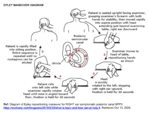
Treatment for BPPV is an otolith repositioning maneuver, which can be performed within a few minutes.
If the patient has a positive Dix-Hallpike (posterior canal BPPV), the treatment is an Epley (or similar) maneuver.
Epley Maneuver Video: Dr. Peter Johns’ Youtube video on how to properly perform an Epley maneuver can be accessed here: https://www.youtube.com/watch?v=7ZgUx9G0uEs
If Dix-Hallpike is negative or atypical, revisit the history and examination. Consider whether symptoms have already resolved, nystagmus is subtle (video recording can help record and review eye movements), or another diagnosis better explains the presentation. If the history and general ED neuro screening exam are otherwise reassuring and consistent with BPPV, even with a negative or atypical Dix-Hallpike, it may be reasonable to still trial a repositioning maneuver (eg. Epley) in select patients.
For the less common horizontal canal BPPV (positive supine roll test), the repositioning maneuver is called a Gufoni maneuver, but this is a much less common form of BPPV, a more nuanced diagnosis to make, and a more finicky repositioning test, so it would warrant referencing your resources and/or deferring to a vestibular physiotherapist if appropriate.
Clinical pearl: Disposition is more than naming the diagnosis. Consider symptom severity, baseline function, frailty and fall risk, ability to mobilize safely, the likelihood of a central or systemic cause, and whether follow-up and return precautions are reliable. Give explicit instructions to return for new focal neurologic symptoms, severe sudden headache, new neck pain, worsening symptoms, or other concerning changes. Imaging is NOT routinely recommended for a typical episodic positional presentation without red flags, but investigate or consult according to clinical concern and local pathways.
Summary: A Simplified 4-Step Approach to EPISODIC Vertigo
STEP 1: Identify Red Flags. Screen for abnormal vital signs, focal neurologic deficits, dangerous Ds, new hearing or visual field deficits, central nystagmus at rest, severe new headache/neck pain, significant trauma, and inability to sit/stand/walk unaided. Consider systemic causes.
STEP 2: Establish Timing & Triggers. Clarify onset, duration of the intense symptoms, whether symptoms persist at rest, what provokes them, and how the patient feels between attacks. Classify the presentation as generally episodic or continuous.
STEP 3: Perform the Appropriate Vertigo Test. For episodic, position-triggered symptoms without spontaneous nystagmus at rest, use the Dix-Hallpike test. Do not use HINTS Plus as a routine test for episodic positional vertigo (we’ll review more about HINTS Plus in the Vertigo Pt 2 episode on continuous vertigo, where HINTS Plus testing comes into play).
STEP 4: Treat & Disposition. Treat confirmed posterior canal BPPV with an Epley maneuver. If the diagnosis is uncertain, broaden the differential, investigate when indicated, and provide safe disposition and explicit return precautions. Routine neuroimaging is NOT indicated or helpful unless you are worried about central TIA or vertebral artery dissection features and/or risk factors on history/physical exam.
Key Clinical Take-Home Pearls for EPISODIC Vertigo
- Do NOT overly rely on the patient’s word choice/descriptor alone (eg. “spinning,” “light-headed,” or “off balance”) to rule diagnoses in or out. Clarify timing, triggers, associated symptoms, and what the patient was doing at onset.
- Symptomatic worsening with movement does NOT distinguish peripheral from central vertigo. Dizziness from any cause typically worsens with movement.
- Start with a focused but thorough neurologic screening exam and don’t forget to assess gait. New inability to sit, stand, or walk unaided is concerning for a central cause and should not be dismissed.
- Choose the right vertigo test depending on whether the vertigo is episodic or continuous. Choosing the wrong test will lead you astray. For brief, triggered episodes without spontaneous nystagmus at rest, use positional testing (Dix Hallpike) and NOT HINTS testing to assess.
- A characteristic positive Dix-Hallpike test supports posterior canal BPPV. Treat with an otolith-repositioning maneuver such as Epley rather than routinely prescribing vestibular suppressants.
- If BPPV cannot be confidently diagnosed, keep the differential open and base investigation and disposition on the whole clinical picture, not a single negative test.
Bottom Line
A careful history, a deliberate neurologic screen, and an observed gait assessment are the foundation of the ED vertigo evaluation. For episodic positional symptoms, the key is to recognize BPPV, confirm it with the appropriate positional test, and treat it with a repositioning maneuver.
Key References
- Edlow et al. Guidelines for reasonable and appropriate care in the emergency department 3 (GRACE-3): Acute dizziness and vertigo in the emergency department. Acad Emerg Med. 2023;30:442-486.
- Puissant et al. Approach to Acute Dizziness/Vertigo in the Emergency Department: Selected Controversies Regarding Specialty Consultation. Stroke. 2024;55(10):2584-2588.
- Edlow et al. A New Diagnostic Approach to the Adult Patient with Acute Dizziness. J Emerg Med. 2018 Apr;54(4):469-483.
- Kaski et al. The International Classification of Vestibular Disorders: Achievements, challenges, and future directions. J Vestib Res. 2025;35(3):105-112.
- Wang et al. ACR Appropriateness Criteria Dizziness and Ataxia: 2023 Update. J of Amer Coll of Rad. 2024;21:S100-S125.
- Dr. Peter Johns & Dr. Scott Weingart. Spin Class Vertigo Course. Accessible here: https://emneuro.com/spinclass/
- Dr. Katie Lin. SYNAPSE: EM Neuro Essentials Course. Accessible here: https://synapsecourse.com/
Dr. Elisha Targonsky has no conflicts of interest to declare. Dr. Peter Johns is the co-creator for the Spin Class Vertigo Course (https://emneuro.com/spinclass/). Dr. Katie Lin is the creator of the SYNAPSE: EM Neuro Essentials Course (https://synapsecourse.com/).



Leave A Comment