ECG Cases – Making complexes simple is a monthly blog by Jesse McLaren (@ECGcases), a Toronto emergency physician with an interest in emergency cardiology quality improvement and education. Each post features a number of ECGs related to a particular theme or diagnosis (with a focus on acute coronary occlusion), so you can test your interpretation skills. We challenge you with missed or delayed diagnosis, those with false positive diagnosis, and those that had a rapid and correct diagnosis. Cases are followed by a quick summary of the literature that relates to the cases, and we bring it home with practice changing pearls that you can use on your next shift.
Share your interesting ECGs with us!
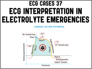
ECG Cases 37 ECG interpretation in electrolyte emergencies
While most of us have a clear algorithm in our minds for the management of life-threatening hyperkalemia, the same may not be said about the other life-threatening electrolyte abnormalities. In this ECG Cases blog Dr. Jesse MacLaren gives us an approach to potassium, calcium and magnesium abnormalities including risk factor assessment, ECG interpretation and management pearls...
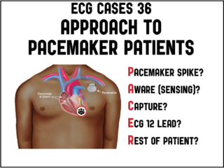
ECG Cases 36 – PACER Mnemonic for Approach to Pacemaker Patients
In this month's ECG Cases blog Dr. McLaren explains the PACER mnemonic approach to patients with pacemakers: Pacemaker spike: is it appropriately presence/absent, is there pacemaker-mediated tachycardia (apply magnet) or is there failure to pace (apply magnet to stop sensing, cardio consult)? Aware (sensing): is it normal, is there oversensing (underpacing: apply magnet) or undersensing (treat reversible causes, cardio consult). Capture: if there are pacemaker spikes is there capture, or failure to capture (treat reversible causes, cardio consult). ECG 12 lead: are there signs of hyperkalemia (extra wide QRS, peaked T) or Occlusion MI (Modified Sgarbossa Criteria) that need immediate treatment. Rest of patient: is there a complication of pacemaker insertion related to the pocket (hematoma, infection), lead (pneumothorax, DVT), or heart (pericardial perforation), or is there an emergency unrelated to the pacemaker (eg dehydration, sepsis, GI bleed)...
ECG Cases 35 – ECG Approach to Takotsubo Syndrome
Takotsubo Syndrome is usually triggered by an emotional or physical stress leading to acute catecholaminergic myocardial stunning. The initial ST elevation phase of Takotsubo Syndrome mimics Occlusion MI, can not be distinguished by patient factors or POCUS findings, and requires immediate angiogram. The subsequent phase of Takotsubo Syndrome has T wave inversion in an apical distribution, which can mimic reperfusion, but often has very deep T wave inversions and a very long QT interval. Takotsubo Syndrome is a retrospective diagnosis of exclusion—with an angiogram ruling out occlusion, a ventriculogram showing apical ballooning, and a follow up echo showing recovery of LV function. Complications of Takotsubo Syndrome include LV failure, apical thrombus, and polymorphic VT from long QT. Jesse McLaren guides us through 10 ECGs to elucidate these important take home points...
ECG Cases 34 – ECG Interpretation in Aortic Dissection
Which patients with ECG evidence of coronary occlusion require a CT scan to rule out aortic dissection? What are the range of ECG findings in acute aortic dissection and how do they change management? Dr. Jesse McLaren guides us through 9 cases to answer these and other questions on ECG interpretation in aortic dissection...
ECG Cases 33 Brugada Syndrome: 3-Step Approach to Diagnosis and Management
Jesse McLaren guides us through 7 cases and explains his 3-step approach to diagnosing and managing Brugada syndrome in this month's ECG Cases blog...
ECG Cases 32 Prehospital ECG pearls and pitfalls
In this ECG Cases blog we review 8 cases of patients with prehospital ECGs and explore prehospital ECGs for diagnosing STEMI, Occlusion MI, false STEMI, code STEMI, dynamic ischemic changes, truncated voltages. Can you avoid the pitfalls and spot the pearls that help to make the diagnosis?


