

Epistaxis is one of those presentations that ranges from a minor nuisance to a true airway and hemorrhage emergency. Most nosebleeds are anterior and stop with simple measures, yet the dangerous posterior bleed may present with little blood coming from the nares while the patient is swallowing, coughing, vomiting, hypoxic and quietly losing a significant amount of blood. So how do we approach epistaxis in a way that is efficient for the common bleed, but aggressive enough for the rare life-threatening one? In this episode with guest experts Dr. Leeor Sommer and Dr. Doron Sommer, we tackle the questions that come up at the bedside: How can you tell an anterior bleed from a posterior bleed before you can actually see the source? What is the correct way to position and compress the nose—and why do so many patients do it wrong? Which topical vasoconstrictor should you use, and where does topical tranexamic acid fit given the conflicting evidence? What is the best approach when you cannot identify a discrete bleeding vessel: absorbable hemostatic agents, a Rapid Rhino, Merocel, or something else? How do you recognize that an anterior pack has failed and it is time to move to posterior packing? What is the safest way to place a dual-balloon posterior pack or Foley catheter? In the patient with torrential posterior epistaxis, when should you secure the airway—and how do you intubate when the oropharynx is filling with blood and your video laryngoscope camera may be useless? Should IV TXA be given in severe epistaxis? Does treating the blood pressure actually help stop the bleeding? Do patients with nasal packing need prophylactic antibiotics? Who can safely go home, who needs ENT urgently, and who needs admission or monitored care? And perhaps most importantly, what are the simple technical details that make the difference between a nosebleed that stops in 10 minutes and one that turns into a 3 a.m. disaster? And many more…
Podcast: Play in new window | Download (Duration: 1:39:57 — 91.6MB)
Subscribe: Apple Podcasts
Podcast production, sound design & editing by Anton Helman; Voice editing by Braedon Paul
Written Summary and blog post by Anton Helman, September 2026
Cite this podcast as: Helman, A. Sommer, D. Sommer, L. Epistaxis Management. Emergency Medicine Cases. September, 2026. https://emergencymedicinecases.com/epistaxis-management. Accessed October 19, 2026
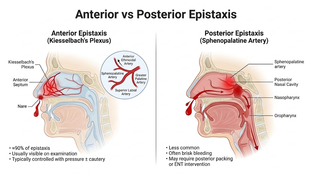
About 90-95% of epistaxis is anterior. The clinically important anterior territory is the anterior septum, particularly Kiesselbach’s plexus, where the majority of bleeds arise and where a culprit vessel can often be seen after adequate vasoconstriction, suction and lighting. Some “anterior” bleeds arise laterally from the inferior turbinate or hide around a septal deviation, making them much harder to visualize.
Posterior epistaxis accounts for approximately 5–10% of cases and commonly arises from branches of the sphenopalatine artery, a terminal branch of the internal maxillary artery. These bleeds tend to be brisker, more difficult to visualize with anterior rhinoscopy, and more likely to require packing or advanced hemostatic intervention. Importantly, failure to visualize a source does not establish a posterior bleed; clot, inadequate vasoconstriction, poor visualization or altered anatomy may obscure an anterior source. The pattern of blood flow provides useful clues but is not definitive. Blood predominantly exiting the anterior nares while the patient leans forward favors an anterior source, whereas persistent posterior pharyngeal bleeding, particularly with little anterior drainage, should raise suspicion for posterior epistaxis and its associated risks of significant hemorrhage and airway compromise.
Predictors of challenging epistaxis management
- Anticoagulants and antiplatelet agents: DOACs, warfarin and dual antiplatelet therapy.
- Hematologic disease or impaired hemostasis: von Willebrand disease, hemophilia, thrombocytopenia, uremia, hereditary hemorrhagic telangiectasia and liver disease.
- Prior recurrent or posterior epistaxis.
- Dry or traumatized mucosa: chronic intranasal steroids or decongestants, home oxygen, CPAP without adequate humidification, dry air, frequent nose blowing/picking and occupational dust exposure.
- Altered anatomy: prior facial trauma or surgery, head and neck cancer/radiation, septal deviation or septal perforation.
- Vascular comorbidity and frailty, including hypertension, CKD and diabetes.
Triage red flags: when epistaxis is a resuscitation case
The sick epistaxis patient usually declares themselves through airway contamination, abnormal physiology or the bleeding pattern. Do not be reassured by a preserved blood pressure: younger and physiologically robust patients may compensate with tachycardia until late. Do not dismiss tachycardia as anxiety in a patient with a history of substantial blood loss.
| Airway | Hemodynamics | Bleeding pattern |
| Blood pooling in posterior pharynx; choking/coughing; gurgling; hypoxia; recurrent hematemesis; altered mental status. | Tachycardia out of proportion to anxiety; presyncope/orthostasis; cool clammy skin; history of large-volume blood loss. | Bilateral bleeding at onset; brisk flow directly into throat; pulsatile/high-volume bleeding; no visible anterior source; failure of proper compression/topicals/anterior packing. |
General overview of management of anterior epistaxis
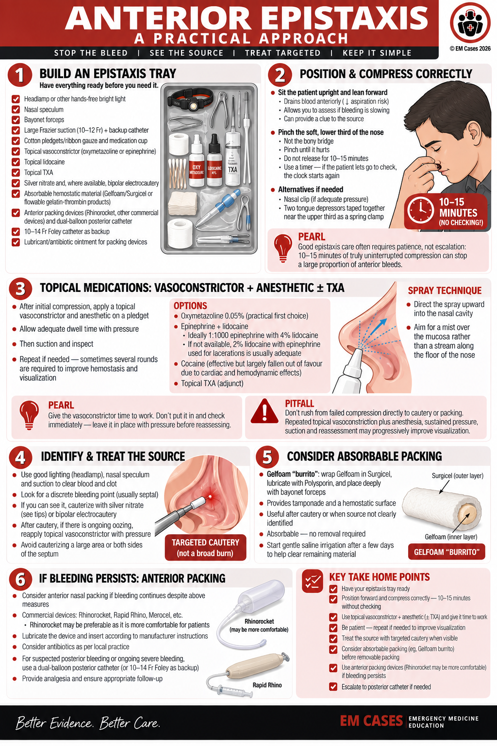
Step 1: Build an epistaxis tray before you need it
A standardized setup prevents the common failure mode of hunting for equipment while the nose continues to bleed. Useful items include:
- Headlamp or other hands-free bright light; nasal speculum; bayonet forceps.
- Large enough Frazier suction (about 10–12 Fr) with a backup catheter because clot can obstruct it.
- Cotton pledgets or ribbon gauze and a small medication cup.
- Topical vasoconstrictor (eg, oxymetazoline or epinephrine) and topical lidocaine.
- Topical TXA, silver nitrate and, where available, bipolar electrocautery.
- Absorbable hemostatic material such as Gelfoam/Surgicel or flowable gelatin-thrombin products.
- Anterior balloon/tampon devices and a dual-balloon posterior catheter; a 10–14 Fr Foley as backup.
- Lubricant/antibiotic ointment for appropriate packing devices.
Dr. Leeor Sommer Anterior Epistaxis Management video
https://youtu.be/Mj3yrIldKd4
Step 2: Position forward and compress correctly
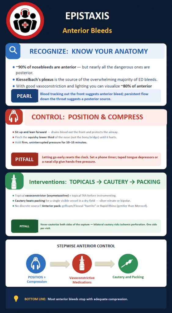
Initial management of uncomplicated epistaxis should begin with upright positioning, forward head tilt and firm, sustained nasal compression. Leaning forward allows blood to drain anteriorly rather than accumulating in the posterior pharynx, reducing swallowing, nausea and potential airway contamination. Compression should be applied to the soft lower third of the nose, below the nasal bones, thereby directly compressing the anterior septal vasculature. The AAO-HNS clinical practice guideline recommends sustained compression for at least 5 minutes, while emergency medicine reviews commonly recommend 10–15 minutes of uninterrupted pressure. Manual pressure or an appropriately positioned nasal clip may be used. Importantly, pressure should not be intermittently released to assess for hemostasis; adequate duration of continuous compression should be allowed before escalating to cautery or packing. Explain to the person applying pressure that if they let go, they need to start the clock again!
Pearl: There is evidence to suggest that placing ice in the mouth against the hard palate decreases blood from to the nose and may improve time to epistaxis cessation
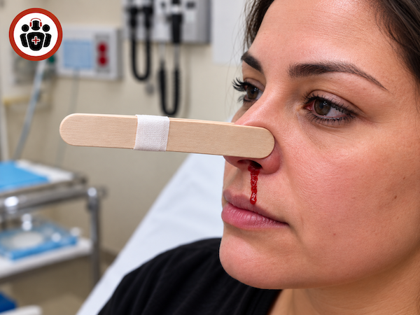
Pearl: Two tongue depressors taped together near their upper third can be used as a simple spring clamp when sustained manual pressure is difficult or the standard nasal clamp provides inadequate pressure.
Step 3: Topical epistaxis medications: vasoconstrictor + anesthetic ± TXA
After sustained compression, the next step is to apply a topical vasoconstrictor combined with local anesthetic, followed by another period of sustained pressure. Epinephrine combined with lidocaine (see below) is one option favored by our experts, as well as oxymetazoline 0.05% or xylometazoline combined with lidocaine. Cocaine provides both vasoconstriction and anesthesia but has largely fallen out of favour because of its cardiac and hemodynamic effects. For epinephrine, mix topical epinephrine with topical lidocaine and applying it using thin cotton, ribbon gauze or gauze placed into the nasal cavity with bayonet forceps.
- Epinephrine: 1:1,000 topically
- Lidocaine: 4% lidocaine, specifically the topical/intubation spray
If 1:1000 epinephrine and 4% lidocaine spray are not available, 2% lidocaine with epinephrine typically used for lacerations may be adequate.
The key is to give the vasoconstrictor time to work. Leave the topical medication in place with pressure before returning to reassess rather than putting it in and checking almost immediately. If the first attempt does not provide adequate control or visualization, repeat the vasoconstrictor/topical anesthetic application—sometimes several times—because each round may improve hemostasis and visualization enough to identify a discrete vessel that can then be cauterized.
Pearl: Oxymetazoline or xylometazoline spray technique: direct the spray upward into the nasal cavity rather than along the floor of the nose, aiming to create a good mist over the mucosa rather than simply shooting a stream posteriorly.
Pitfall: Don’t rush from failed compression directly to cautery or packing. Repeated topical vasoconstriction plus anesthesia, sustained pressure, suction and reassessment may progressively improve visualization until the bleeding vessel becomes apparent and amenable to definitive cautery.
Topical TXA is attractive because systemic absorption is minimal and the safety profile is good, but the efficacy literature is mixed. Several smaller studies and meta-analyses suggested faster hemostasis or less packing, whereas the larger NoPAC randomized trial did not reduce the need for anterior packing when TXA was used as the studied intervention. Our experts still consider it reasonable as part of a topical “trifecta” with vasoconstrictor and anesthetic, rather than as a replacement for compression and source control.
Floseal and Surgiflo are flowable hemostatic agents made from a Gelfoam/thrombin combination. These tools can be particularly useful for brisk bleeding, anticoagulated patients or abnormal anatomy where a balloon has nothing reliable to compress against — for example, a large septal perforation or post-radiation/post-surgical anatomy. They need to be pressed against the bleeding surface and into the nooks and crannies, rather than simply injected into the nasal cavity, because active bleeding can push the material away. Their major drawback is cost, so they are generally reserved for situations where their particular advantages are worthwhile.
Step 4: Identify and cauterize the source of epistaxis
Visualization is the rate-limiting step. Give the topical agents time to work — sometimes more than one round — then remove clot with suction. If you can identify one discrete vessel in a relatively dry field, cautery is preferable to reflexive packing. Silver nitrate works by chemical cautery and is much less effective when brisk blood immediately washes it away. Use a brief, focused application to a small area rather than painting a large segment of septum.
Tips to improve visualization of the bleeding source
- Use a headlamp so both hands remain free for the nasal speculum, suction, forceps and treatment.
- Use a nasal speculum and good suction—preferably a larger Frazier suction that is less likely to become obstructed by clot.
- Clear blood and clot before looking for the source. Have more than one suction available because clot can quickly obstruct it.
- Topicalize first, then look. Apply a vasoconstrictor plus topical anesthetic on cotton, ribbon gauze or gauze and allow it time to work with compression before re-examining the nose.
- Be patient and repeat the process. A second or even third round of vasoconstrictor/topical anesthetic may progressively improve visualization rather than immediately escalating to packing.
- Gentle irrigation can help when blood and clot repeatedly obscure the field
- Look for a very discrete bleeding point. The goal is to get the field dry enough to identify the actual vessel so that cautery can be targeted rather than applied broadly.
- Remember that not every difficult-to-see bleed is posterior. Some anterior bleeds arise laterally around the inferior turbinate or around a corner and can be difficult to visualize without an endoscope.
- Account for abnormal anatomy. A deviated septum, septal perforation, previous surgery, radiation or resection may make the usual bleeding sites difficult to see and require a different approach.
Tips for using silver nitrate cautery
- Be very focal with the silver nitrate. Think of it as a small “spot weld” directly on the culprit vessel rather than broadly cauterizing the surrounding septal mucosa.
- If it starts oozing after silver nitrate, don’t keep cauterizing. Place cotton/gauze soaked with a topical vasoconstrictor directly over the area and have the patient apply pressure.
- Give the silver nitrate time to work. Silver nitrate produces a chemical burn; brisk bleeding can wash over the area before the cautery has had an opportunity to take effect. Vasoconstriction and pressure help prevent this.
- Avoid cauterizing corresponding areas on both sides of the septum, because opposing mucosal injuries increase the risk of septal injury/perforation.
Pitfall: Avoid cauterizing opposing sides of the septum at the same visit. Bilateral septal cautery can compromise cartilage blood supply and increase the risk of septal perforation.
Pearl: Once you’ve used silver nitrate to cauterize the culprit bleeding source, add some topical 1:1000 epinephrine afterwards to minimize the chance of re-bleeding.
Bipolar electrocautery: effective but painful
Where available, bipolar electrocautery can be more effective than silver nitrate and can work in a slightly wetter field. Topical anesthesia may be insufficient; a small local injection of lidocaine with epinephrine at the target may be required. Use a low setting appropriate to the device (roughly 10–20 W), keep the bipolar tips separated, and apply for only 1–2 seconds at a time.
Step 5: Anterior epistaxis packing: absorbable before removable when possible
When cautery fails or no discrete vessel can be found, packing is next. A patient-centered goal is to avoid sending patients home with a removable nasal pack whenever feasible: removable packs are uncomfortable, may require analgesia and create a second procedure when they are removed. Absorbable packing has the advantage of causing minimal pain and does not require follow-up for removal.
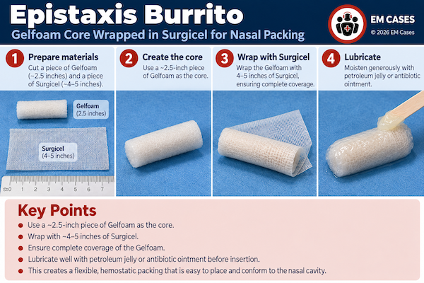
The EM Cases ‘epistaxis gelfoam burrito’
The Gelfoam “burrito” is an absorbable packing technique that can be used after cautery or when a bleeding source cannot be clearly identified, before moving to removable nasal packing favored by our experts. A piece of Gelfoam is wrapped in Surgicel, creating a small “burrito,” lubricated with Polysporin and then placed deeply into the nasal cavity with bayonet forceps so that it sits against the bleeding area. The combination provides both tamponade and a hemostatic surface while avoiding the discomfort and subsequent removal required with traditional nasal packing. Because the material is absorbable and gradually dissolves, it can be left in place, with gentle saline irrigation started after a few days to help clear the remaining material.
Step 6: Removable anterior epistaxis packs
If a removable pack is required, balloon packs such as Rapid Rhino are often easier to insert, better tolerated and allow titration of pressure compared with rigid tampon sponges. Soak a Rapid Rhino in water for 10–30 seconds to activate its hydrogel coating. Insert it fully — the external marker should be at the nostril — because a partially inserted balloon will preferentially inflate outside the nose where resistance is lowest. Inflate only enough to achieve hemostasis and tolerable pressure.
Pitfall: A nasal balloon that is sticking out of the nostril is not just uncomfortable — it may be mechanically ineffective because the balloon expands where resistance is lowest.
General overview of posterior epistaxis management
Recognizing posterior epistaxis and when to escalate
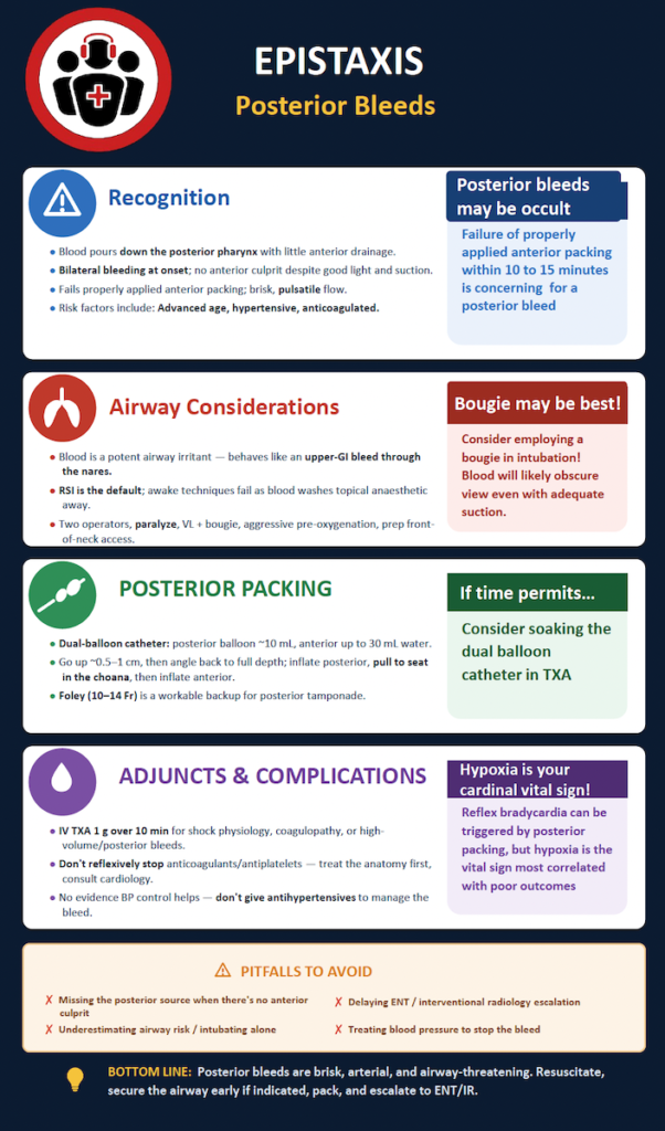
Posterior epistaxis is a clinical diagnosis. Think posterior when:
- Blood primarily pours into the posterior pharynx with little anterior drainage,
- Bleeding is bilateral from the outset,
- No anterior source is seen despite proper suction/vasoconstriction/lighting, or
- A correctly placed anterior pack fails after 10–15 minutes.
- High-volume pulsatile hemorrhage and the older anticoagulated patient increase concern.
- Persistent oropharyngeal bleeding despite anterior measures,
- Hemodynamic compromise/significant transfusion requirement
- Anatomy that makes anterior control impossible
Dr. Leeor Sommer’s EM Cases Summit Posterior Epistaxis Management Video
https://youtu.be/cGEE3aSF43g
Massive posterior epistaxis airway management: Treat it like a contaminated airway
Blood is a potent airway irritant and an enemy of laryngoscopy. It obscures the camera, clogs suction and can trigger coughing, vomiting, aspiration and laryngospasm. The patient with persistent hypoxia, blood that cannot be cleared from the oropharynx, altered mental status or massive bleeding with shock physiology needs an airway plan early — ideally with anesthesia/airway backup and ENT simultaneously mobilized.
In torrential bleeding, awake techniques are often impractical because topical anesthesia is washed away and the patient may not have the time or cooperation required. RSI is generally the default approach according to our experts. Use two skilled airway operators, prepare front-of-neck access, preoxygenate as aggressively as possible, and have large-bore suction (Decanto) immediately available. Video laryngoscopy is reasonable, but assume the camera may become useless. A bougie may be particularly valuable when blood obscures the view. A rapidly placed nasal pack before laryngoscopy may reduce ongoing inflow if it can be done without delaying airway control.
Pearl: In a blood-filled airway, the bougie may become your best visual aid: prepare for the camera to fail and have a second skilled operator dedicated to suction.
Placement of commercially available dual-balloon catheter for posterior epistaxis
A dedicated dual-balloon catheter is preferred over a Foley catheter because the anterior balloon anchors the device.
- Lubricate the dual-balloon catheter before insertion. It is a double-balloon device with separate posterior and anterior balloons.
- Start by directing the catheter slightly upward as it enters the nostril for the first 0.5cm-1cm, following the initial orientation of the nasal vestibule.
- Then angle the catheter posteriorly, directing it toward the back of the head rather than continuing upward.
- Advance the catheter all the way to the hilt. The goal is to advance it sufficiently posteriorly that the tip and posterior balloon have passed beyond the choana.
- Inflate the posterior balloon with water. The transcript describes using roughly 10 mL of water in the posterior balloon.
- Pull the catheter anteriorly until the posterior balloon seats firmly against the choana. This is the critical maneuver that creates posterior tamponade and applies pressure to the posterior bleeding area.
- Inflate the anterior balloon with water. It can accommodate up to 30 mL, but the transcript emphasizes that less is usually required—inflate according to what is needed and what the patient can tolerate.
The anterior balloon serves two purposes: it helps hold the posterior balloon securely against the choana without requiring an external clip, and it simultaneously provides anterior nasal tamponade if there is also an anterior bleeding source.
If a dedicated device is unavailable, a 10–14 Fr Foley can provide posterior tamponade, but it requires secure external fixation and meticulous padding to avoid pressure necrosis of the ala. Posterior packing is a bridge, not definitive therapy; involve ENT early.
Posterior packing complications
Posterior packs can worsen oxygenation by forcing mouth breathing and reducing effective airway cross-section. Historical teaching emphasizes reflex bradycardia, but clinically significant bradycardia from the pack itself appears uncommon. Hypoxia, ongoing hemorrhage and pressure injury are the practical threats. Overinflation or excessive external tension can cause alar or septal necrosis.
Pearl: After posterior packing, oxygen saturation is a cardinal vital sign. Do not let concern about reflex bradycardia distract from hypoxia and ongoing hemorrhage.
Tranexamic acid in severe epistaxis
For the critically ill patient with hemorrhagic shock physiology, a high-volume/posterior bleed or systemic coagulopathy, our experts recommend considering TXA 1 g IV over 10 minutes as an adjunct. This should never delay mechanical source control, resuscitation or definitive ENT/IR management.
Anticoagulants and antiplatelets: source control first!
Antithrombotic therapy can make a nosebleed harder to stop, but the first treatment remains local hemostasis. Do not reflexively stop aspirin, dual antiplatelet therapy or a DOAC after a controlled anterior bleed. The thrombotic indication matters: a mechanical valve, recent thromboembolism or high-risk coronary disease may make interruption hazardous. Conversely, the patient in extremis with uncontrolled life-threatening hemorrhage may require anticoagulant reversal. For the difficult middle ground, involve the clinician who can best quantify thrombotic risk — cardiology, hematology or neurology — and make the decision jointly. A useful discharge step is to explicitly tell patients whose antithrombotic therapy should continue not to stop it on their own.
Pitfall: A controlled nosebleed is not, by itself, a reason to stop a well-indicated antithrombotic medication; unnecessary interruption may trade epistaxis for stroke, valve thrombosis or coronary thrombosis.
Definitive therapy of posterior epistaxis may ultimately require endoscopic arterial control or interventional radiology embolization.
Hypertension and epistaxis: treat the bleed, not the number
Hypertension is associated with epistaxis, but is generally not a cause of epistaxis. A high ED blood pressure is often amplified by pain, anxiety and adrenergic stress. There is no evidence that acutely lowering blood pressure is a hemostatic treatment for otherwise uncomplicated epistaxis. Control the bleeding, relieve distress, then reassess. Persistent hypertension can be addressed according to usual hypertension principles and followed longitudinally.
Antibiotics with nasal packing: not routine
The literature does not support routine systemic prophylactic antibiotics for most short-duration anterior nasal packs. Consider antibiotics selectively when packing will remain for more than 48–72 hours, with ribbon gauze, in immunocompromised patients, with pre-existing sinus infection or other particularly high-risk circumstances. Lubricating non-balloon packs with topical antibiotic ointment and arranging timely removal is sufficient for most patients.
Epistaxis disposition, aftercare and ENT follow-up
A patient with a controlled anterior bleed can usually go home after a period of observation if there is no continued blood around the pack or down the throat, vital signs are stable, the airway is secure and follow-up is reliable. Removable anterior packs generally need reassessment/removal after about 48 hours. Removal in a setting capable of treating rebleeding is prudent, particularly for anticoagulated patients.
For the first three days after hemostasis or cautery, advise patients to leave the nose alone: no picking, vigorous blowing or unnecessary manipulation. Thereafter, humidification and gentle moisturization can reduce recurrent crusting. Patients with recurrent severe or unilateral bleeds, repeated ED visits, new change in their bleeding pattern, significant coagulopathy, posterior packing or packing complications need early ENT follow-up and often endoscopic assessment.
Posterior packs generally require admission. A young, otherwise healthy patient with bleeding controlled by a dedicated posterior pack may not always require ICU, but frailty, cardiopulmonary disease, substantial blood loss, ongoing oxygen requirement or concern for rebleeding should push toward a closely monitored setting.
Key take home points for ED epistaxis management
- Start with the patient upright and leaning forward, then compress the soft lower third of the nose continuously for 10–15 minutes.
- Do not rush past vasoconstriction, anesthesia, suction and lighting. The better you can see, the more likely you are to cauterize a discrete source and avoid packing.
- Use absorbable hemostatic material before a removable pack when appropriate; if a removable pack is needed, balloon devices are often easier to titrate and better tolerated.
- Suspect posterior epistaxis when blood preferentially runs down the throat, bleeding is bilateral/brisk, no anterior source is found, or a properly placed anterior pack fails.
- Massive posterior epistaxis is an airway emergency. Hypoxia, gurgling, inability to clear blood and altered mental status should trigger early airway help, aggressive suction planning and preparation for a contaminated RSI.
- Posterior packing is a bridge to definitive care. Watch oxygenation closely and escalate early to ENT/IR when bleeding is uncontrolled.
- Topical TXA has mixed evidence; it is reasonable as an adjunct with vasoconstrictor/anesthetic. Consider IV TXA in selected critically ill high-volume/posterior bleeds, but never instead of source control.
- Do not reflexively stop antiplatelet or anticoagulant therapy after a controlled bleed, and do not acutely treat hypertension simply to stop epistaxis.
- Routine prophylactic systemic antibiotics are not required for most short-duration anterior packs.
- Good discharge instructions — pressure technique attempted for 10 minutes twice with ice in the mouth against the hard palate if bleeding recurs , nasal rest, humidification/moisturization and a clear plan for pack removal/rebleeding — are part of definitive treatment.
Key References
- Seikaly H. Epistaxis. N Engl J Med. 2021;384(10):944-951.
- Tunkel DE, et al. Clinical practice guideline: Nosebleed (Epistaxis). Otolaryngol Head Neck Surg. 2020;162(1_suppl):S1-S38.
- Newton E, Lasso A, Petrcich W, Kilty SJ. An outcomes analysis of anterior epistaxis management in the emergency department. J Otolaryngol Head Neck Surg. 2016;45:24.
- Bellew SD, et al. Effect of intranasal vasoconstrictors on blood pressure: a randomized, double-blind, placebo-controlled trial. J Emerg Med. 2018;55(4):455-464.
- McLeod RWJ, et al. Intranasal cautery for the management of adult epistaxis: systematic review. J Laryngol Otol. 2017;131(12):1056-1064.
- Lange JL, Peeden EH, Stringer SP. Are prophylactic systemic antibiotics necessary with nasal packing? A systematic review. Am J Rhinol Allergy. 2017;31(4):240-247.
- Joseph J, Martinez-Devesa P, Bellorini J, Burton MJ. Tranexamic acid for patients with nasal haemorrhage (epistaxis). Cochrane Database Syst Rev. 2018;12:CD004328.
- Reuben A, et al. The use of tranexamic acid to reduce the need for nasal packing in epistaxis (NoPAC): randomized controlled trial. Ann Emerg Med. 2021;77(6):631-640.
- Zahed R, et al. Topical tranexamic acid compared with anterior nasal packing in patients taking antiplatelet drugs: a randomized controlled trial. Acad Emerg Med. 2018;25(3):261-266.
- Min HJ, Kang H, Choi GJ, Kim KS. Association between hypertension and epistaxis: systematic review and meta-analysis. Otolaryngol Head Neck Surg. 2017;157(6):921-927.
- Corrales CE, Goode RL. Should patients with posterior nasal packing require ICU admission? Laryngoscope. 2013;123(12):2928-2929.
- Marks PLG, Parks AR. Suction-assisted airway catheter insertion for airway management in severe epistaxis: a case report. CJEM. 2024;26(4):283-285.
Drs. Helman and Dr. Leeor Sommer have no conflicts of interest to declare. Dr. Doron Sommer reports relationships with Sanofi, GSK and AstraZeneca through clinical research and advisory board activities; Stryker as a consultant (Stryker produces HemoPore, an absorbable chitosan hemostatic agent); and Medtronic through speaking engagements.




Leave A Comment