

Imaging decisions are part of the daily work of Emergency Medicine—but when to image, when to wait, and what to do when the ideal test isn’t available are often far more nuanced than they might appear. How do we balance the diagnostic power of CT against cumulative radiation risk? How much should cumulative radiation exposure influence our decision to order another CT? In suspected appendicitis, when is it safe to wait until morning for ultrasound rather than obtain a CT overnight? Can the emerging traumatic brain injury biomarkers GFAP and UCH-L1 help safely avoid CT, particularly in rural and remote settings? And in suspected cauda equina syndrome, can CT lumbar spine help risk stratify patients when MRI isn’t available? In this episode, Anton is joined by Dr. Amit Shah, academic, community and rural Emergency Physician and Associate Professor at Western University, to examine the evidence behind these everyday imaging dilemmas and help us choose the right test, for the right patient, at the right time…
Podcast: Play in new window | Download (Duration: 48:36 — 44.6MB)
Subscribe: Apple Podcasts
Podcast production, sound design & editing by Anton Helman
Written Summary and blog post by Anton Helman August, 2026
Cite this podcast as: Helman, A. Shah, A. Imaging decisions in Emergency Medicine. Emergency Medicine Cases. August, 2026. https://emergencymedicinecases.com/imaging-decisions-emergency-medicine. Accessed October 25, 2026
How concerned should we be about radiation from CT?
In patients being considered for CT—particularly younger patients and those with multiple previous CT examinations—how should the potential long-term radiation-associated cancer risk influence the decision to image?
CT is an extraordinarily useful diagnostic tool, but its use has increased substantially. In 2023, approximately 93 million CT examinations were performed on 61.5 million patients in the United States. One study suggests that radiation from these CTs could ultimately contribute to approximately 103,000 future cancers, potentially representing approximately 5% of annual cancer diagnoses if current utilization and radiation-dose patterns continue.
Key study: Smith-Bindman et al, 2025
P: US patients undergoing CT examinations in 2023
I: Exposure to ionizing radiation from contemporary CT imaging
C: Modeled background lifetime cancer incidence without CT-associated radiation exposure
O: Projected lifetime radiation-induced cancers
The investigators used CT utilization data together with organ-specific radiation doses and National Cancer Institute risk models. Of the approximately 103,000 projected cancers, about 91% were attributable to CT examinations performed in adults, largely because adults undergo far more CT examinations despite children having greater individual radiation susceptibility. Abdomen/pelvis CT accounted for the largest proportion of projected CT-associated cancers in adults.
These numbers require careful interpretation. They represent modeled future risk rather than observed cancers causally attributed to individual CT examinations, and the precise magnitude of low-dose radiation risk remains uncertain. Nonetheless, the study reinforces an important principle: when millions of CT examinations are performed, even a small individual risk may translate into meaningful population harm.
Radiation risk is not uniform
Radiation-associated cancer risk is strongly influenced by age, sex, body region and scan protocol. Younger patients have substantially greater lifetime attributable risk because of greater tissue radiosensitivity and longer remaining lifespan during which a radiation-induced malignancy could develop. This becomes particularly relevant in patients with recurrent presentations—renal colic, chronic abdominal pain and recurrent headache, for example—who may accumulate numerous CT examinations over years.
The practical implication is not that CT should be avoided when indicated. Rather, previous imaging should become part of the imaging history. Before ordering another CT, review the patient’s prior imaging whenever feasible and ask:
- What is my pretest probability of important pathology?
- Will CT change immediate management?
- Has the patient already undergone multiple CT examinations?
- Could ultrasound, observation, serial examination or follow-up reasonably answer the clinical question?
- Is the risk of delaying imaging greater than the potential harm of imaging?
When the probability of a dangerous diagnosis is very low, time and serial examination are legitimate diagnostic tools. Observation with repeat vital signs, repeat examination and explicit follow-up may sometimes provide more value than immediate imaging.
=> Do not fear CT when the patient needs CT. But do not treat CT as harmless. The younger the patient and the greater the cumulative exposure, the stronger the rationale for considering observation or non-ionizing alternatives when clinically appropriate.
Suspected appendicitis: CT tonight or ultrasound in the morning?
In a clinically stable patient with suspected appendicitis when CT is immediately available but ultrasound is delayed several hours, how urgently is definitive imaging required?
Sometimes, concern prompting immediate CT is that delaying definitive imaging could lead to uncomplicated appendicitis progressing to perforation. However, appendicitis rarely progresses from uncomplicated inflammation to perforation over a few hours. Duration of symptoms appears more important than a short in-hospital delay.
Key study: Jiang et al, 2021
P: Adults undergoing appendectomy for acute appendicitis
I: Increasing duration from symptom onset to surgery
C: Shorter symptom duration
O: Appendiceal perforation and operative outcomes
In this prospective cohort of 255 patients, the incidence of perforation increased progressively with symptom duration. Compared with patients with 24–48 hours of symptoms, symptom duration of ≥48 hours was independently associated with perforation (OR 4.64; 95% CI 1.76–12.27). This suggests that symptom duration should influence imaging urgency. A clinically well patient early in the course of illness may reasonably wait until the morning for ultrasound when that strategy avoids CT radiation. Conversely, prolonged symptoms—particularly beyond approximately 48 hours—should increase concern for complicated disease prompting urgent CT. Other concerning features include evolving peritonitis, hemodynamic instability, significant fever and marked inflammatory abnormalities.
Should empiric antibiotics be given while awaiting imaging?
For a clinically stable patient with suspected but unconfirmed uncomplicated appendicitis, antibiotics given solely to prevent progression to perforation are not clearly beneficial.
The PERFECT-Antibiotics randomized trial provides useful evidence.
Key study: Jalava et al, 2025 — PERFECT-Antibiotics
P: Adults with presumed uncomplicated acute appendicitis awaiting appendectomy
I: Cefuroxime plus metronidazole while awaiting surgery
C: No antibiotics while awaiting surgery; both groups received perioperative prophylaxis
O: Appendiceal perforation at surgery
Among 1,774 patients, perforation occurred in 8.3% with antibiotics versus 8.9% without antibiotics, meeting the prespecified criterion for noninferiority of withholding antibiotics. Importantly, this applies to patients with presumed uncomplicated appendicitis; patients with suspected perforation, generalized peritonitis, significant fever or other indications for urgent treatment were excluded.
=> A stable patient early in the course of suspected uncomplicated appendicitis does not necessarily require CT simply because ultrasound is unavailable for several hours. Clinical trajectory, duration of symptoms and features suggesting complicated disease should determine urgency. Empiric antibiotics solely to prevent perforation while awaiting confirmation have not been shown to reduce perforation in uncomplicated appendicitis.
Can serum biomarkers help avoid CT after mild traumatic brain injury?
Can serum biomarkers safely help rule out significant traumatic intracranial injury in patients who cannot otherwise be cleared clinically?
The Canadian CT Head Rule remains the starting point for adult minor head injury. In the original prospective cohort of 3,121 patients, the high-risk criteria were 100% sensitive for the need for neurological intervention. However, decision rules intentionally favour sensitivity. This creates a particularly difficult problem in remote environments where satisfying a CT criterion may mean air transport over hundreds of miles. Two blood biomarkers—glial fibrillary acidic protein (GFAP) and ubiquitin C-terminal hydrolase-L1 (UCH-L1)—have emerged as potential adjuncts.
Key study: Bazarian et al, 2021
P: Adults with nonpenetrating mild/moderate traumatic brain injury undergoing head CT
I: Rapid plasma GFAP/UCH-L1 testing using the i-STAT platform
C: Head CT as reference standard
O: Detection/rule-out of acute traumatic intracranial injury
Among 1,901 patients with GCS 13–15, the rapid biomarker test demonstrated:
Sensitivity: 95.8%
Specificity: 40.4%
Negative predictive value: 99.3%
Positive predictive value: 9.8%
Their poor specificity means they should not replace clinical assessment or established CT decision rules. If indiscriminately applied to low-risk patients, false-positive results could actually increase imaging. The potential role for these biomarkers is more targeted according to our expert: patients who cannot be cleared by a validated decision rule but in whom the absolute probability of clinically significant intracranial injury remains relatively low, particularly when CT requires difficult or resource-intensive transport.
=> Apply validated clinical decision rules first. GFAP/UCH-L1 testing may become a useful second-stage rule-out strategy in selected patients, particularly where CT access is difficult. A positive biomarker test is not diagnostic of intracranial hemorrhage, and the test should not become an indiscriminate screening test for all minor head injuries.
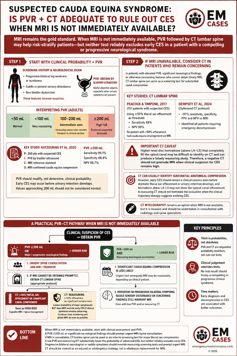
Suspected cauda equina syndrome: Is PVR + CT adequate to rule out CES when MRI is not immediately available?
When MRI is not immediately available, can post-void residual (PVR) and CT lumbar spine be combined to risk-stratify patients with suspected cauda equina syndrome (CES)?
MRI remains the gold-standard imaging test for CES, but obtaining an emergency MRI may require prolonged delays or transfer. In this setting, PVR followed selectively by CT lumbar spine may provide a practical risk-stratification pathway in selected patients—with the important caveat that neither test reliably excludes early CES in a patient with a compelling or progressive neurological syndrome.
Step 1: Start with clinical probability and PVR
History and neurological examination for CES screening come first. Progressive bilateral leg weakness or numbness, saddle or perianal sensory disturbance and new bladder dysfunction should increase suspicion. PVR provides a useful objective adjunct, particularly when urinary symptoms are present. PVR <50mL is considered normal. PVR <100 mL is very reassuring, ≥200 mL is the evidence-supported high-risk threshold, and 100–200 mL represents an intermediate zone where increasing residual volume should raise suspicion and be interpreted in the context of the neurological examination.
Key study: Katzouraki et al, 2020
P: 260 patients referred with suspected CES
I: PVR measured by bladder ultrasound
C: MRI reference standard
O: MRI-confirmed cauda equina compression
A PVR ≥200 mL had 94.1% sensitivity, 66.8% specificity and 98.7% NPV for cauda equina compression. Thus, ≥200 mL is a useful threshold for identifying higher-risk patients, while a low PVR substantially reduces the probability of significant compression. However, PVR should modify rather than determine clinical probability. A patient may have early CES before developing urinary retention, and surgical intervention during the early phase of illness is more likely to portend a good outcome. Similarly, values approaching 200 mL should not be regarded as definitively normal simply because they fall below the study threshold.
Step 2: If MRI is unavailable, consider CT in patients who remain concerning
For patients with an elevated PVR, significant neurological findings or otherwise concerning clinical features who cannot obtain timely MRI, lumbar CT can serve as a screening test for substantial canal compromise.
Key studies: Peacock and Timpone, 2017; Dempsey et al, 2024
In 151 patients with suspected CES, using ≥50% thecal sac effacement as the CT threshold produced 98% sensitivity and 99% NPV for significant spinal stenosis. No patient with <50% effacement had cauda equina impingement on MRI. More recently, Dempsey et al evaluated an optimized CT protocol and reported approximately 97% sensitivity, specificity, PPV and NPV compared with MRI for compressive disc pathology; importantly, CT identified all patients requiring emergency decompression.
Important CT caveat: Higher-level disc herniations (above L4–L5) that completely fill the spinal canal may be more difficult to identify on CT and can produce a falsely reassuring study. Thus, a negative CT should not preclude MRI when clinical suspicion for CES remains high.
=> CT can usually identify substantial anatomical compression. However, early CES should remain a clinical concern even before dramatic thecal sac effacement or urinary retention develops, and disc herniations above L4-L5 may not show the typical fecal effacement of CES compared to lower lumbar levels. Therefore, a reassuring CT should not terminate the evaluation when the clinical trajectory strongly suggests evolving CES.
CT myelography remains another potential option when MRI is not available, but it is invasive and should be undertaken in consultation with radiology and spine specialists.
A practical PVR–CT pathway when MRI is not immediately available
Clinical suspicion of CES → obtain PVR
PVR ≥200 mL or major/progressive neurological findings → HIGHER RISK
→ Urgent spine consultation and MRI/transfer.
→ If MRI cannot be obtained promptly, obtain CT lumbar spine using an appropriate protocol as an interim screening test.
→ ≥50% thecal sac effacement or significant canal compromise: treat as high risk and expedite MRI/spine management.
→ Reassuring CT: lowers the probability of major compression but does not exclude early CES if the clinical syndrome remains concerning.
PVR <200 mL + reassuring neurological examination → LOWER RISK
→ Significant cauda equina compression is less likely.
→ Depending on the clinical picture, urgent rather than emergent MRI may be reasonable.
→ Persistent or progressive bilateral symptoms, saddle sensory disturbance or other concerning findings still warrant MRI despite a low PVR.
The key is to think of PVR and CT as sequential probability modifiers rather than binary rule-out tests. PVR helps identify patients more likely to have clinically important compression; CT can then identify substantial anatomical canal compromise when MRI is inaccessible. Neither replaces MRI when clinical suspicion remains high.
=> When cauda equina syndrome is on the differential, start with clinical assessment and PVR. A PVR ≥200 mL or significant neurological findings should prompt urgent MRI/spine consultation. If MRI is unavailable, CT lumbar spine can be used as an interim screening test for major thecal sac compression. A low PVR and reassuring CT substantially lower the probability of advanced CES, but neither reliably excludes early CES. Progressive bilateral neurological or saddle symptoms should therefore override reassuring screening tests and prompt urgent MRI. CT should be viewed as an adjunct or contingency strategy, not a wholesale replacement for MRI.
Putting it together: Imaging is a clinical intervention
Imaging decisions are often framed as though ordering a test is the conservative choice and withholding one is the risky choice. That framing is incomplete. Every imaging decision represents a balance between competing risks: missed or delayed diagnosis on one side, and radiation, incidental findings, downstream testing, transport, resource utilization and prolonged ED stays on the other.
3 principles of imaging decisions in Emergency Medicine
- Pretest probability matters more than the availability of the scanner. A readily available CT does not necessarily make CT the correct test.
- Time is a diagnostic tool. Serial examination, observation and planned reassessment can sometimes resolve uncertainty without radiation.
- Context matters. The threshold for imaging or transfer may appropriately differ between an urban ED with immediate CT and MRI access and a remote nursing station where obtaining imaging requires prolonged transport.
Clinical decision rules, biomarkers, bedside tests and alternative imaging modalities can refine probability, but none eliminates the need for clinical judgment. The goal is not to order fewer CTs. The goal is to order the right imaging, in the right patient, at the right time.
References
- Smith-Bindman R, Chu PW, Azman Firdaus H, et al. Projected lifetime cancer risks from current computed tomography imaging. JAMA Intern Med. 2025;185(6):710-719. doi:10.1001/jamainternmed.2025.0505.
- Jiang X, Namba Y, Takayama T, et al. Does the time from symptom onset to surgery affect the outcomes of patients with acute appendicitis? A prospective cohort study of 255 patients. Asian J Endosc Surg. 2021;14:361-367. doi:10.1111/ases.12870.
- Jalava K, Sallinen V, Lampela H, et al. Role of preoperative antibiotic treatment while awaiting appendectomy: the PERFECT-Antibiotics randomized clinical trial. JAMA Surg. 2025;160(7):745-754. doi:10.1001/jamasurg.2025.1212.
- Stiell IG, Wells GA, Vandemheen K, et al. The Canadian CT Head Rule for patients with minor head injury. Lancet. 2001;357(9266):1391-1396. doi:10.1016/S0140-6736(00)04561-X.
- Bazarian JJ, Welch RD, Caudle K, et al. Accuracy of a rapid glial fibrillary acidic protein/ubiquitin carboxyl-terminal hydrolase L1 test for the prediction of intracranial injuries on head computed tomography after mild traumatic brain injury. Acad Emerg Med. 2021;28(11):1308-1317. doi:10.1111/acem.14366.
- Katzouraki G, Zubairi AJ, Hershkovich O, Grevitt MP. A prospective study of the role of bladder scanning and post-void residual volume measurement in improving diagnostic accuracy of cauda equina syndrome. Bone Joint J. 2020;102-B(6):677-682. doi:10.1302/0301-620X.102B6.BJJ-2020-0195.R1.
- Peacock JG, Timpone VM. Doing more with less: diagnostic accuracy of CT in suspected cauda equina syndrome. AJNR Am J Neuroradiol. 2017;38(2):391-397. doi:10.3174/ajnr.A4974.
- Dempsey PJ, Ryan DT, Lambe G, et al. An optimized CT protocol for detecting suspected cauda equina syndrome: a comparative analysis with MRI. AJNR Am J Neuroradiol. 2024;45(9):1370-1377. doi:10.3174/ajnr.A8315.
Drs. Helman and Shah have no conflicts of interest to declare
Now test your knowledge with a quiz.



Leave A Comment