

Topics in this EM Quick Hits podcast
Amna Karabegovic on a case of pediatric altered LOA and neurocritical care review (2:09)
Anand Swaminathan on management of pulmonary hypertension and RV failure (24:32)
Brit Long on cytokine release syndrome (30:44)
Andrew Tagg on management of pediatric distal radius fractures (39:31)
Jesse McLaren on ECG interpretation for the bradycardic patient (46:05)
Sara Gray & Katie Lin on Coaching the EM Mind Part 2: stress management and optimizing safety on shift (52:55)
Podcast: Play in new window | Download (Duration: 1:39:00 — 90.7MB)
Subscribe: Apple Podcasts
Podcast production, editing and sound design by Anton Helman
Podcast content, written summary & blog post by Sara Brade, edited by Anton Helman, July, 2026
Cite this podcast as: Helman, A. Lin, K. Karabegovic, A. Swaminathan, A. Long, B. Tagg, A. McLaren, J. Gray, S. EM Quick Hits 72 – EMC²: Peds Neurocritical Care, Pulmonary Hypertension, Cytokine Release Syndrome, Peds Wrist Fracture, Bradycardia ECG Interpretation, Coaching the EM Mind Part 2 Emergency Medicine Cases. July, 2026. https://emergencymedicinecases.com/em-quick-hits-72-july-2026/ . Accessed October 11, 2026.
EM Cases Cases (EMC²): Pediatric Neurocritical Care
Rapid neurologic assessment and differential diagnosis for the altered pediatric patient
In the crashing child suspected of neurologic catastrophy, focus on the essential neurologic exam: eyes/pupils, verbal response and motor response.
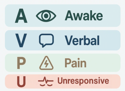
AVPU—Alert, responds to Voice, responds to Pain, Unresponsive—can provide a fast common language when calculating an exact GCS is impractical, and correlates well with GCS.
- Organize the differential into structural versus metabolic causes. Check glucose, temperature and vital signs immediately, but maintain a low threshold for urgent head CT when the clinical picture points towards a structural emergency (i.e. headache, vomiting and markedly depressed consciousness).
- Appropriate CT should not be delayed solely because the patient is a child who is at risk from radiation exposure when the presentation is life-threatening and the result will change immediate management.
Immediate management priorities in a child with raised intracranial pressure (ICP)
- Elevate the head of the bed and keep the neck midline; provide adequate analgesia and antiemetic therapy.
- Maintain oxygenation, normothermia, euglycemia and euvolemia with isotonic fluid. Avoid hypoxia and hypotension, both of which worsen secondary brain injury.
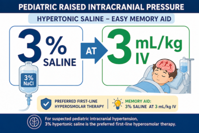
- For suspected pediatric intracranial hypertension, 3% hypertonic saline is the preferred first-line hyperosmolar therapy: an easy memory aid is 3% saline at 3 mL/kg IV.
- Mannitol remains an alternative when hypertonic saline is unavailable, but monitor closely for osmotic diuresis, hypovolemia and electrolyte abnormalities.
Pediatric neurocritical airway management considerations
- Aim for first-pass success with the most experienced available operator, video laryngoscopy, full preoxygenation and a clearly communicated backup plan.
- Minimize hypoxemia and hemodynamic swings during airway management. Laryngoscopy-related bradycardia in children is often driven by hypoxia; preventing desaturation is more important than routine atropine pretreatment.
- Have atropine and epinephrine immediately available. Atropine is not routinely required for pediatric RSI, but may be considered in selected infants (age <1 yr) or when significant bradycardia is already present.
- Use age-specific blood pressure ranges, avoid hypotension and seek pediatric ICU guidance for hypertension. Evidence-based BP targets for spontaneous pediatric ICH remain limited. The priorities are to avoid hypotension and maintain cerebral perfusion while generally avoiding blood pressure above approximately the 95th percentile for the child’s age, sex and size.
Inhaled nitroglycerine for pulmonary hypertension and RV failure
Patients with acute pulmonary hypertension—from a large pulmonary embolism or decompensated chronic pulmonary arterial hypertension—can deteriorate rapidly when right-ventricular preload falls or RV afterload rises. The immediate goal is to improve oxygenation and ventilation while avoiding interventions that precipitate RV collapse.
Initial respiratory support of patients with pulmonary hypertension in the ED
- Correct hypoxemia and hypercarbia because both increase pulmonary vascular resistance. Avoid endotracheal intubation when a reasonable alternative exists. Transition to invasive positive-pressure ventilation can reduce venous return, increase RV afterload and trigger abrupt hemodynamic collapse.
- Bi-level non-invasive ventilation (BPAP) may improve oxygenation and lower CO2 while preserving some spontaneous negative-pressure breathing. High-flow nasal oxygen is reasonable when hypoxemia is the main issue, but it will not reliably correct substantial hypercarbia.
Inhaled nitroglycerine as pulmonary vasodilator in the ED for pulmonary hypertension
- Inhaled nitric oxide is a classic rapid pulmonary vasodilator, but it may not be immediately available in the ED. Inhaled epoprostenol is another option, although pharmacy preparation and delivery can introduce clinically important delays.
- A practical bridge is using nebulized nitroglycerin while waiting for inhaled nitric oxide or epoprostenol. Nitroglycerin is rapidly converted into nitric oxide in the lungs. The suggested dose is 5 mg by nebulizer every 5 minutes. Practically, it is difficult to fit 5mg into the nebulizer chamber so our expert suggests filling the chamber with nitroglycerin and increasing flow rate to max. This is an off-label, time-critical strategy that should be aligned with local critical-care, respiratory-therapy and pharmacy protocols.
EM Cases Crit Cases 7 – Pulmonary Hypertension – A Fine Balance
Recognition and management of Cytokine Release Syndrome after CAR T-cell therapy
Cytokine Release Syndrome (CRS) is a severe systemic inflammatory response caused by a rapid surge of cytokines. It is increasingly encountered after chimeric antigen receptor T-cell therapy and may range from a mild influenza-like illness to shock, hypoxic respiratory failure and multiorgan dysfunction. Other triggers include: monoclonal antibodies, haploidentical donor stem cell transplantation, and nonprotein cancer drugs.
CAR T-cell therapy uses genetically modified T cells to attack malignant cells and it is used to treat cancers like ALL, CLL, and multiple myeloma. Examples include:
-
- Tisagenlecleucel (KYMRIAH)
- Axicabtagene ciloleucel (YESCARTA)
- Brexucabtagene autoleucel (TECARTUS)
- Lisocabtagene maraleucel (BREYANZI)
- Idecabtagene vicleucel (ABECMA)
- Ciltacabtagene autoleucel (CARVYKTI)
Consider CRS when a patient within two weeks of CAR T-cell therapy has a fever of at least 38°C that is not better explained by another cause, together with constitutional symptoms, hypotension or hypoxia. There is no single diagnostic ED laboratory or imaging test. Evaluate simultaneously for sepsis, tumour lysis syndrome, pulmonary embolism, heart failure and treatment-related organ injury.
ED management of CRS
- Resuscitate with judicious fluids, supplemental oxygen and vasopressors as required. Provide antipyretics. Evaluate and treat end-organ dysfunction.
- Because sepsis may be indistinguishable at presentation, give broad-spectrum antibiotics when the patient is significantly ill or infection remains plausible.
- Contact the treating oncologist or cellular-therapy team early, regardless of severity of illness.
- Treatment with tocilizumab, an IL-6 receptor antagonist may be considered under specialist guidance.
- Severe or refractory CRS may require corticosteroids.
Bottom line: A recent CAR T-cell infusion (within last 2 weeks) plus otherwise unexplained fever, hypotension or hypoxia should immediately raise CRS. Resuscitate, cover possible infection and involve the oncology/cellular-therapy team early—specific therapy is time-sensitive.
Management of pediatric displaced distal radius fractures: the CRAFFT Trial
A completely displaced pediatric wrist looks alarming and traditionally prompts reduction under sedation or general anesthesia, sometimes with fixation. The CRAFFT trial asked whether immediate anatomic correction improves outcomes that matter to children and families—or whether casting alone allows sufficient remodeling.
The paper: Perry DC, Zimmermann A, Achten J, et al. Non-surgical casting versus surgical reduction for children with severely displaced distal radial fractures (the CRAFFT Study): a multicentre, randomised, controlled non-inferiority trial and economic evaluation. The Lancet. Published online April 8, 2026.
PICO:
Population: 750 children aged 4–10 years with severely displaced distal radial fractures across 49 UK hospitals; 44% had completely off-ended fractures. Intervention: casting without purposeful manipulation, general anesthesia or procedural sedation. Comparison: surgical (closed or open) reduction under general anesthesia or conscious sedation, with fixation permitted at the surgeon’s discretion. Primary outcome: parent-reported PROMIS upper-extremity function at three months. The prespecified non-inferiority margin was −2.5 points.
Results:
-
- At three months, surgery produced a small functional advantage: adjusted mean difference 1.64 PROMIS points in favour of reduction.
- The confidence interval crossed the conservative non-inferiority margin, so casting did not technically demonstrate non-inferiority for the primary analysis.
- The observed difference was below the magnitude families considered meaningful enough to justify surgery, and there was no functional difference by six or twelve months.
- Early procedural complications—including pressure injury, wound infection, scarring and nerve irritation—occurred mainly in the surgical group. Refracture was slightly more frequent with casting but remained uncommon overall.
- Casting saved approximately £1,665 per patient and had a 100% probability of being cost-effective at the UK thresholds studied.
Limitations and application:
-
- Treatment was unblinded and the primary outcome was parent reported, so expectation may have influenced early scores.
- The pragmatic design allowed variation in treatment technique and crossover, although this also improves real-world relevance.
- The clearest application is to children aged 4–10 years. Results should not be extrapolated automatically to older children approaching skeletal maturity.
- The trial does not support cast-first care when there is neurovascular compromise, threatened skin, an open or intra-articular fracture, or another reason for urgent reduction.
- Implementation should be collaborative with local orthopedic teams because practice patterns and thresholds for intervention vary.
Bottom line: For most children aged 4–10 years with a severely displaced distal radius fracture, an intact neurovascular exam and no skin threat, a cast-first strategy is a reasonable default—even when the X-ray looks dramatic. Treat the child’s function and safety, not the radiograph alone.
Bradycardia and the 12-lead ECG: A systematic approach and BRADI mnemonic
ACLS is designed to stabilize primary electrical bradyarrhythmias, but many ED patients are bradycardic because of a secondary, reversible process. The rhythm strip tells you what the rate and rhythm are; the history, vital signs and full 12-lead ECG may tell you why.
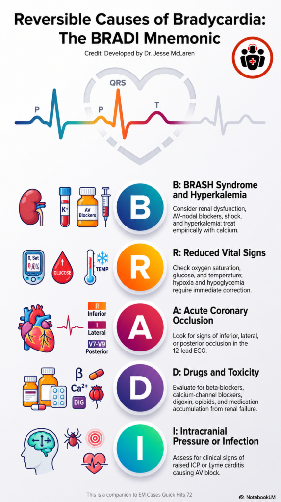
The BRADI mnemonic for reversible causes of bradycardia in the ED
B – BRASH syndrome and hyperkalemia: Think renal dysfunction, AV-nodal blockers, shock and hyperkalemia. Hyperkalemic bradycardia may be sinus or junctional and may show PR prolongation, QRS widening, axis change or peaked T waves. Atropine and pacing may fail; treat suspected hyperkalemia promptly, including empiric calcium when the clinical and ECG picture fits.
R – Reduced vital signs: Check the complete set: oxygen saturation, glucose and temperature. Hypoxia and hypoglycemia require immediate correction. Hypothermia may produce bradycardia, shivering artifact, prolonged intervals and prominent J waves.
A – Acute coronary occlusion: The left circumflex perfuses the SA node as well as the lateral and posterior territories, while the RCA perfuses the SA and AV nodes as well as the inferior and posterior territories. In a patient with ischemic symptoms and bradycardia, look for signs of inferior, lateral or posterior occlusion: inferior ST elevation or hyperacute T waves, reciprocal change in aVL, or anterior ST depression reciprocal to posterior injury.
D – Drugs: Consider beta-blockers, calcium-channel blockers, digoxin, opioids and medication accumulation from renal failure. Management may require antidotal or supportive therapy, withholding the culprit drug and allowing clearance. Digoxin may produce a shortened QT interval and scooped ST depression, but most drug causes are identified primarily by history.
I – Intracranial pressure (raised) or infection: Raised ICP is primarily a clinical diagnosis, although diffuse deep T-wave inversion and QT prolongation may occur. Lyme carditis may cause high-grade or complete AV block and is a reversible reason to avoid an unnecessary permanent pacemaker.
Bottom line: Stabilize the unstable patient, but do not stop at atropine, epinephrine or pacing. Ask why the patient is bradycardic and use the 12-lead ECG most actively when hyperkalemia or acute coronary occlusion is plausible.
For examples of the BRADI mnemonic, ECG Cases 58.
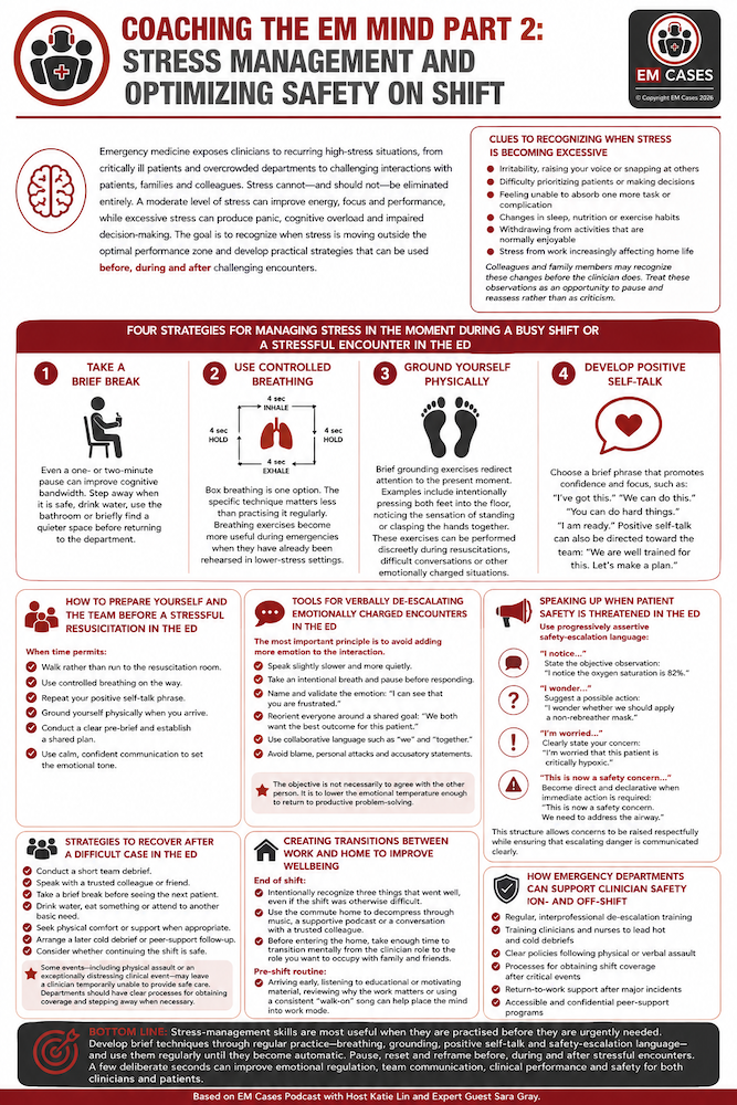
Coaching the EM Mind Part 2: Stress management and optimizing safety on shift

Emergency medicine exposes clinicians to recurring high-stress situations, from critically ill patients and overcrowded departments to challenging interactions with patients, families and colleagues. Stress cannot—and should not—be eliminated entirely. A moderate level of stress can improve energy, focus and performance, while excessive stress can produce panic, cognitive overload and impaired decision-making. The goal is to recognize when stress is moving outside the optimal performance zone and develop practical strategies that can be used before, during and after challenging encounters.
Clues to recognizing when stress is becoming excessive for emergency clinicians
Clinicians may not immediately recognize that they have entered a state of cognitive or emotional overload. Warning signs include:
- Irritability, raising your voice or snapping at others
- Difficulty prioritizing patients or making decisions
- Feeling unable to absorb one more task or complication
- Changes in sleep, nutrition or exercise habits
- Withdrawing from activities that are normally enjoyable
- Stress from work increasingly affecting home life
Colleagues and family members may recognize these changes before the clinician does. Treat these observations as an opportunity to pause and reassess rather than as criticism.
Four strategies for managing stress in the moment during a busy shift or a stressful encounter in the ED
- 1Take a brief break – Even a one- or two-minute pause can improve cognitive bandwidth. Step away when it is safe, drink water, use the bathroom or briefly find a quieter space before returning to the department.
- 2Use controlled breathing – Box breathing is one option. The specific technique matters less than practising it regularly. Breathing exercises become more useful during emergencies when they have already been rehearsed in lower-stress settings.
- Ground yourself physically – Brief grounding exercises redirect attention to the present moment. Examples include intentionally pressing both feet into the floor, noticing the sensation of standing or clasping the hands together. These exercises can be performed discreetly during resuscitations, difficult conversations or other emotionally charged situations.
- Develop positive self-talk – Choose a brief phrase that promotes confidence and focus, such as: “I’ve got this.” “We can do this.” “You can do hard things.” “I am ready.” Positive self-talk can also be directed toward the team: “We are well trained for this. Let’s make a plan.”
How to prepare yourself and the team before a stressful resuscitation in the ED
When time permits:
- Walk rather than run to the resuscitation room.
- Use controlled breathing on the way.
- Repeat your positive self-talk phrase.
- Ground yourself physically when you arrive.
- Conduct a clear pre-brief and establish a shared plan.
- Use calm, confident communication to set the emotional tone
Tools for verbally de-escalating emotionally charged encounters in the ED
The most important principle is to avoid adding more emotion to the interaction.
Practical strategies include:
- Speak slightly slower and more quietly.
- Take an intentional breath and pause before responding.
- Name and validate the emotion: “I can see that you are frustrated.”
- Reorient everyone around a shared goal: “We both want the best outcome for this patient.”
- Use collaborative language such as “we” and “together.”
- Avoid blame, personal attacks and accusatory statements.
The objective is not necessarily to agree with the other person. It is to lower the emotional temperature enough to return to productive problem-solving.
Speaking up when patient safety is threatened in the ED
Use progressively assertive safety-escalation language:
- “I notice…”
State the objective observation: “I notice the oxygen saturation is 82%.” - “I wonder…”
Suggest a possible action: “I wonder whether we should apply a non-rebreather mask.” - “I’m worried…”
Clearly state your concern: “I’m worried that this patient is critically hypoxic.” - “This is now a safety concern…”
Become direct and declarative when immediate action is required: “This is now a safety concern. We need to address the airway.”
This structure allows concerns to be raised respectfully while ensuring that escalating danger is communicated clearly.
Strategies to recover after a difficult case in the ED
The effects of a stressful resuscitation may continue long after the event is over. Immediate recovery strategies include:
- Conduct a short team debrief.
- Speak with a trusted colleague or friend.
- Take a brief break before seeing the next patient.
- Drink water, eat something or attend to another basic need.
- Seek physical comfort or support when appropriate.
- Arrange a later cold debrief or peer-support follow-up.
- Consider whether continuing the shift is safe.
Some events—including physical assault or an exceptionally distressing clinical event—may leave a clinician temporarily unable to provide safe care. Departments should have clear processes for obtaining coverage and stepping away when necessary.
Creating transitions between work and home for emergency physicians to improve wellbeing
- At the end of a shift, intentionally recognize three things that went well, even if the shift was otherwise difficult. Use the commute home to decompress through music, a supportive podcast or a conversation with a trusted colleague.
- Before entering the home, take enough time to transition mentally from the clinician role to the role you want to occupy with family and friends.
- A pre-shift routine can provide the opposite transition. Arriving early, listening to educational or motivating material, reviewing why the work matters or using a consistent “walk-on” song can help place the mind into work mode.
Deep dive on pre-shift strategies: Episode 134 Shift Preparation: Pre-gaming with Rob Orman
How emergency departments can support clinician safety on- and off-shift
Stress management should not be treated solely as an individual responsibility. Emergency departments should consider:
- Regular, interprofessional de-escalation training
- Training clinicians and nurses to lead hot and cold debriefs
- Clear policies following physical or verbal assault
- Processes for obtaining shift coverage after critical events
- Return-to-work support after major incidents
- Accessible and confidential peer-support programs
Bottom line: Stress-management skills are most useful when they are practised before they are urgently needed. Develop brief techniques through regular practice—breathing, grounding, positive self-talk and safety-escalation language—and use them regularly until they become automatic. Pause, reset and reframe before, during and after stressful encounters. A few deliberate seconds can improve emotional regulation, team communication, clinical performance and safety for both clinicians and patients.
None of the authors have any conflicts of interest to declare

Leave A Comment