


In this ECG Cases blog we review 10 patients with possible AV block: is there AV block, and if so where is it, what’s the cause, and what’s the treatment?
Written by Jesse McLaren; Peer Reviewed and edited by Anton Helman. November 2026
10 patients presented with possible AV node block. Is there AV node block and if so, where’s the block, why is it happening, and what’s the treatment?
Case 1: 50 year old with weakness
–
Case 2: 50 year with palpitations
–
Case 3: 75 year old with weakness and dizzy
–
Case 4: 65 year old with palpitations
–
Case 5: 60 year old with presyncope
–
Case 6: 85 year old, history of atrial fibrillation on beta-blocker, with weakness
–
Case 7: 75 year old, history of atrial flutter on beta-blocker, with weakness
–
Case 8: 85 year old, history of chronic kidney disease, with weakness
–
Case 9: 30 year old with acute chest pain
–
Case 10: healthy 30 year old, camping 2 months prior, presenting with syncope
–
AV block pitfalls and pearls
AV node blocks are classified by PR interval as first degree (PR delay but no block), second degree (intermittent AV block), and third degree (complete AV block), and second degree is subdivided into Mobitz type 1 (Wenckebach) and type 2 by whether or not the PR interval prolongs before the block. But there are number of pitfalls: looking at the PR interval in isolation can lead to missing or overcalling AV block, and the classification does not emphasize where/why the block is happening or what to do about it.
In the emergency department there are three important questions for AV block:
1. What are the atria doing, and is there AV block?
Before assessing the AV node we need to look at atrial activity, map out sinus activity to see if there’s a block, and consider AV block if there’s AF/AFL with slow response
- If there’s sinus rhythm, is there AV block?
- Missed AV block includes what appears as ‘sinus bradycardia’ but is actually normal sinus (regular P-P at normal rate) with a second P wave superimposed on the T wave
- AV block mimics include blocked PAC (P wave earlier and with different morphology) or prominent U wave (eg hypokalemia)
- If there’s atrial fibrillation or flutter, is there complete AV block?
- Atrial fibrillation with slow and regular ventricular response = third degree AV block with escape rhythm
- Atrial flutter with slow and regular ventricular response but irregular flutter-QRS relationship = third degree AV block with escape rhythm
2. Is the AV block nodal or infranodal?
The multiple types of AV block can be dichotomized by whether they are within the AV node or below the AV node:
- AV nodal block: may be physiologic and respond to atropine, does not require pacemaker, but may have secondary cause
- First degree: regular P-P, fixed long PR without block
- Second degree type 1: regular P-P, progressive PR prolongation leading to AV block (longest PR before block, shortest PR after block)
- AV infranodal block: not physiologic, atropine can worsen, requires a pacemaker if no reversible cause
- Second degree type 2: regular P-P, fixed PR but AV block. If there is 2:1 block it is more likely to be infranodal if there is bundle branch or fascicular blocks
- Third degree: regular P-P, variable PR and AV dissociation. The escape rhythm may be junctional (typically narrow QRS with a rate around 50) or ventricular (typically wide QRS with a QRS around 30).
3. Is there a secondary cause ?
While nodal vs infranodal stratifies primary electrical blocks (with infranodal requiring a pacemaker if no reversible cause), it’s important to assess the patient and ECG for secondary causes regardless of the type of block. Secondary causes of BRADI-cardia/blocks:
- BRASH/hyperkalemia: can cause a variety of rhythm, conduction, axis, and repolarization abnormalities – requires calcium
- Reduced vitals, eg hypothermia (bradycardia, shivering artifact, long PR/QRS/QT) – requires correction and treating underlying cause
- Acute coronary occlusion, especially right coronary artery that perfuses the AV node (eg inferior ST elevation or hyperacute T wave, reciprocal STD/TWI in aVL, anterior STD from posterior extension) – requires cath lab activation
- Drugs, (eg beta blockers, calcium channel blockers, digoxin that can cause ‘regular AF’ from 3rd degree AV block) – requires holding or reversal
- ICP or Infection (eg lyme carditis, requires antibiotics)
Back to the cases
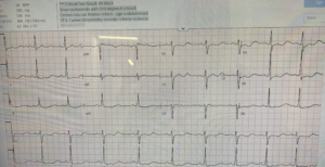
Case 1: 50 year old with weakness
- Heart rate/rhythm: normal sinus rhythm not sinus tachycardia
- Electrical intervals: prominent U wave, not second P wave
- Axis: normal axis
- R-wave progression: pseudo infarct from V1/3 reversal
- Tall/small voltages: normal voltages
- ST/T: diffuse STD
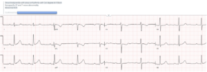
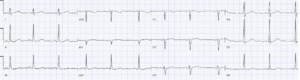
Impression: normal sinus with prominent U wave (not second P wave with AV block as the computer says) and diffuse STD, with weakness = hypokalemia until proven otherwise. Potassium was 1.5. Repeat ECG after potassium correction was normal:
–
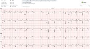
Case 2: 50 year with palpitations
- H: regular P-P then early P wave with different morphology, followed by a pause, then a ventricular escape beat, then back to regular P = sinus with blocked PAC
- E: fixed PR prolongation, normal QRS
- A: left axis from LAFB
- R: delayed R wave
- T: normal voltages
- S: no ST/T changes
Impression: normal sinus with blocked PAC and first degree (nodal) AV block. Labs normal and discharged without treatment
–
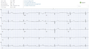
Case 3: 75 year old with weakness and dizzy
- H: normal sinus rhythm (2nd P wave deforms T wave, regular P-P) not sinus bradycardia or blocked PACs
- E: fixed long PR, narrow QRS
- A: normal axis
- R: mild delay
- T: normal voltages
- S: isolated TWI in III
Impression: normal sinus with 2nd degree AV block with 2:1 conduction (could be nodal or infranodal). No reversible causes found, admitted for monitoring and received a pacemaker for ongoing symptomatic AV block.
–
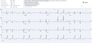
Case 4: 65 year old with palpitations
- H: normal sinus rhythm (regular P-P intervals, no PAC)
- E: fixed PR with every third P wave non-conducted (Mobitz 2 not 1), RBBB
- A: left axis from LAFB
- R: early R wave from RBBB, persisting S wave from LAFB
- T: normal voltages
- S: secondary changes
Impression: normal sinus with Mobitz 2 (infranodal) and bifascicular block. Admitted for pacemaker.
–
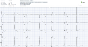
Case 5: 60 year old with presyncope
- H: normal sinus rhythm with regular but dissociated ventricular response
- E: variable not fixed PR (see rhythm strip), wide QRS from RBBB
- A: normal axis
- R: early R from RBBB
- T: normal voltages
- S: inferior Q wave with T wave inversion
Impression: normal sinus with 3rd degree AV block (infranodal) and ventricular escape. Labs were normal, and admitted for a pacemaker.
–
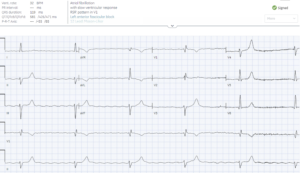
Case 6: 85 year old, history of atrial fibrillation on beta-blocker, with weakness
- H: atrial fibrillation with slow regular ventricular response = escape rhythm
- E: 3rd degree AV block, incomplete RBBB
- A: left from LAFB
- R: early from RBBB
- T: normal
- S: possible peaked T waves
Impression: AF with 3rd degree AV block (infranodal) and ventricular escape rhythm. Not on digoxin, labs normal, and received permanent pacemaker
–
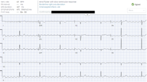
Case 7: 75 year old, history of atrial flutter on beta-blocker, with weakness
- H: atrial flutter with slow and regular ventricular response
- E: variable flutter-to-QRS relationship = third degree AV block with narrow complex QRS
- A: normal axis
- R: late R wave from Q wave V2-3
- T: normal voltages
- S: no ST/T changes
Impression: atrial flutter with third degree AV block (infranodal) and junctional escape. Labs normal, not on digoxin. Admitted for pacemaker.
–
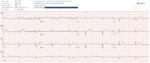
Case 8: 85 year old, history of chronic kidney disease, with weakness
- H: sinus bradycardia
- E: very long and fixed PR interval, normal QRS
- A: left axis from LAFB
- R: delay R wave
- T: low voltages
- S: diffuse peaked T waves (symmetric, pointy, tall relative to QRS)
Impression: sinus bradycardia with first degree AV block (nodal) secondary to hyperkalemia. Initially treated with atropine with no effect. Then potassium returned at 7.1
–
Case 9: 30 year old with acute chest pain
- H: sinus bradycardia
- E: fixed long PR, normal QRS
- A: normal axis
- R: normal R wave
- T: normal voltages
- S: inferior hyperacute T waves (wide based, symmetric, bulky), reciprocal TWI in aVL (concordant but disproportionate to QRS), associated with STD V2-3
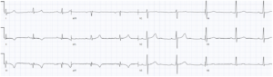
Impression: sinus bradycardia and first degree AV block (nodal) secondary to infero-posterior occlusion MI. Cath lab activated: RCA occlusion. Discharge ECG showed resolution of AV block, and infero-posterior reperfusion T wave inversion (reciprocal ion aVL and 2):
–
Case 10: healthy 30 year old, camping 2 months prior, presenting with syncope
- H: sinus tachycardia with wide complex ventricular escape
- E: variable PR, LBBB morphology
- A: left axis
- R: late R wave typical of LBBB
- T: normal voltages
- S: ST discordant and proportional to QRS (secondary)
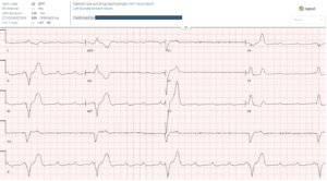
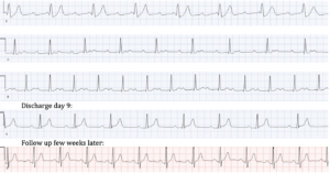
Impression: third degree AV block (infranodal) with ventricular escape, in healthy 30 year old with recent camping = lyme carditis until proven otherwise. Treated empirically with IV ceftriaxone and admitted to hospital. Lyme serology positive, and AV block progressively resolved with antibiotics:
–
Take home points for ECG Cases AV block pitfalls and pearls:
- What are atria doing? Map out P waves to identify subtle second P wave, and differentiate PACs and U waves. Consider AV block if atrial fibrillation with slow and regular ventricular response, and atrial flutter with slow and regular ventricular response but irregular flutter-QRS relationship
- Where’s the block? Is AV block nodal (long PR from first degree, or block preceded by PR prolongation from Mobitz 1) or infranodal (block with fixed PR from Mobitz 2, often with bundle branch block; or variable PR from 3rd degree, with narrow junctional or wide ventricular escape).
- Is there a reversible cause? Is there a secondary BRADI-cardia/block (BRASH/hyperkalemia, Reduced vitals, Acute coronary occlusion, Drugs, ICP or Infection with lyme), or does the patient have a primary electrical block requiring a pacemaker
Further reading: Lee, Gill, Shokr, McLaren. Bradycardia in the emergency department: quality measures and opportunities for improvement. Am J Emerg Med 2026
Elevate your skills! For live, interactive, and CME-accredited courses to elevate your ECG interpretation skills – including a pre-conference course before the 2026 EM Cases Summit, visit www.heartsECGcourse.com




Leave A Comment