

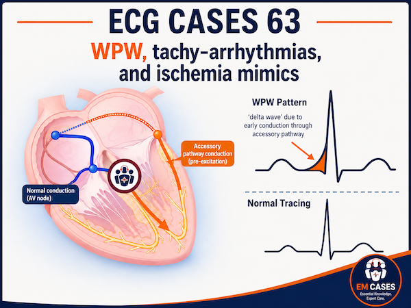
In this ECG Cases blog we review how to identify WPW, treat its associated tachyarrhythmias, and identify its ischemia mimics…
Written by Jesse McLaren; Peer Reviewed and edited by Anton Helman. August 2026
Please consider a donation to EM Cases to ensure ongoing high quality free open access medical education here: https://emergencymedicinecases.com/donation/
5 patients presented with potential WPW. What’s the diagnosis and treatment?
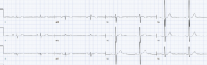
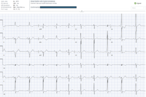
Case 1: 40 year old with episodic palpitations, attributed to anxiety. Old then new ECG
–
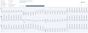
Case 2: 40 year old with recurring palpitations
–
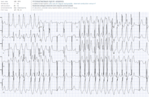
Case 3: 30 year old with palpitations
–
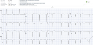
Case 4: 50 year old with epigastric pain and vomiting
–
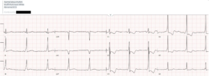
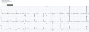
Case 5: 60 year old with two hours of chest pain. Serial ECG
–
WPW, tachyarrhythmias, and ischemia mimics
In 1930 Wolff, Parkinson and White reported described patients with structurally normal hearts who presented with tachy-arrhythmias, whose baseline ECGs had an intermittent pattern of short PR and wide QRS that would fluctuate spontaneously or based on vagal stimulation.[1] Wolff-Parkinson-White (WPW) pattern indicates an accessory pathway allowing ventricular pre-excitation, WPW syndrome is the clinical presentation of associated tach-arrhythmias, and the pattern may also mimic or mask ischemia.
WPW pattern: Intermittent baseline ECG
Normal embryonic development produces a fibrous ring that separates the atria and ventricles, channeling conduction through the AV node into the His-Purkinje fibers. The AV node is like a toll booth before a highway: it causes a pause (normal PR interval) before rapid conduction (narrow QRS). In WPW there’s a congenital defect in the fibrous separation, with a bypass tract that can conduct between atria and ventricles. This accessory pathway is like a gravel road that bypasses the toll booth and highway: without having to pause at the AV node there is early entry into the ventricles (short PR), but as a result of going around the AV node the ventricular depolarization does not have access to the rapid His-Purkinje system and is slow (QRS widened by initial delta wave). Like a stream of traffic, the ECG appearance of WPW reflects the ratio of AV vs accessory pathway conduction, which can change over time including within the same ECG: if all conduction goes does the AV node the ECG will look normal (concealed accessory pathway), but the greater proportion of conduction using the accessory pathway the more obvious the short PR and delta wave that widens the QRS.[2]
WPW syndrome: Symptomatic tachyarrhythmias
The WPW accessory pathway can create a circuit for arrhythmias (atrioventricular reentrant tachycardia, or AVRT, which can go up or down the AV node) or a way for atrial fibrillation to bypass the AV node. WPW syndrome can cause recurring palpitations or syncope, or other symptoms including shortness of breath, weakness or anxiety. During the tachy-arrhythmias it can be difficult to identify the accessory pathway. But the ECG indicates the mechanism of arrhythmia and use of the accessory pathway, which guides treatment, and the post-cardioversion ECG can then look for features of WPW.[2]
- Orthodromic AVRT: uses the AV node to conduct to the ventricles (orthodromic) and the accessory pathway to return (with a RP interval longer than typical ANVRT). Therefore looks like other SVT (regular narrow complex tachycardia) and is treated the same: AV node blockers or electrical cardioversion
- Antidromic AVRT: uses the accessory pathway to conduct to the ventricles and AV node to return. Therefore it looks like monomorphic VT (regular wide complex tachycardia) and is treated the same: procainamide or electrical cardioversion
- AF + WPW: looks like atrial fibrillation, but because the accessory pathway provides uninhibited access to the ventricles the rate is very fast (up to 300), and because the QRS reflects the ratio of AV vs accessory pathway its morphology changes. Can treat with procainamide if stable, but with rates up to 300 it is faster to electrically cardiovert. Avoid AV node blockers (including amiodarone) which will channel more conduction down the accessory pathway
Mimic or mask ischemia
Depending on the specific location of the accessory pathway, it can alter the direction of conduction in the frontal plane (axis deviation) and horizontal plane (abnormal R-wave progression), including exaggerating voltage (pseudo-hypertrophy). The abnormal depolarization (wide QRS) can also cause abnormal repolarization (ST/T waves) that can mimic or mask ischemia.[3] So patients may have incidental WPW that is misdiagnosed as ischemia (including negative delta waves that mimic pathological Q waves except they are preceded by a short PR), or present with Occlusion Myocardial Infarction that is difficult to identify because of the baseline changes. As with other kinds of abnormal depolarization (eg bundle branch block or hypertrophy), secondary repolarization abnormalities are discordant (opposite direction) and proportional to the QRS, which can help identify superimposed primary ischemia.[4-5]
Back to the cases
Case 1: 40 year old with episodic palpitations, attributed to anxiety. Old then new ECG
- Heart rate/rhythm: normal sinus rhythm in both
- Electrical conduction: repeat ECG has intermittent short PR with delta (beats 1-4, 8-9)
- Axis: left axis during pre-excitation only
- R-wave progression: early R wave during pre-excitation only
- Tall/small voltages: tall voltages without LVH
- ST/T: secondary, discordant and proportional ST/T during pre-excitation
= WPW syndrome with intermittent accessory pathway conduction. Referred to electrophysiology clinic.
–
Case 2: 40 year old with recurring palpitations
- H: regular narrow complex tachycardia with long RP (best seen leads V1 and V3)
- E: narrow QRS
- A: normal
- R: abnormal progression
- T: normal voltages
- S: rate related diffuse STD
= orthodromic AVRT vs atypical AVNRT: Modified Valsalva failed, then cardioversed with adenosine:
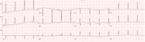
- H: normal sinus
- E: short PR with delta wave
- A: normal
- R: early R wave in V2
- T: normal
- S: mild secondary discordant STD in V3
= WPW syndrome with orthodromic AVRT treated with AV blocker. Referred to electrophysiology clinic
–
Case 3: 30 year old with palpitations
- H: irregularly irregular rhythm (AF) with rates up to 300
- E: variable QRS
- A: variable axis
- R: normal
- T: tall
- S: secondary discordant ST
= WPW syndrome with AF, which is contraindication to AV node blockers (including amiodarone). Electrical cardioversion:
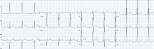
- H: normal sinus
- E: short PR with delta wave (upright in I and inverted in III)
- A: normal
- R: early
- T: pseudo-LVH in lead aVL
- S: pseudo-inferior infarct
= sinus with WPW, with pseudo LVH and pseudo infarct. Follow up ECG post-ablation is normal (except for tall voltages, appropriate for healthy 30 year old):
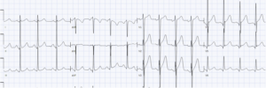
–
Case 4: 50 year old with epigastric pain and vomiting
- H: normal sinus rhythm with PC
- E: short PR with delta wave (eg upright in I/aVL, negative in II/III/aVF)
- A: left axis from pseudo-inferior infarct
- R: late R wave progression, pseudo-anterior infarct
- T: tall voltages in aVL, pseudo-LVH
- S: I/aVL have discordant STD/TWI, and III/aVF have discordant STE and upright T waves
= incidental WPW pattern with pseudo-LVH and pseudo-infarct. Patient diagnosed with bowel obstruction. Troponins and echo normal. ECG was baseline pattern
–
Case 5: 60 year old with two hours of chest pain. Serial ECG
- H: sinus borderline bradycardia in both
- E: short PR + delta wave in first, not second
- A: normal
- R: early R wave in first (pre-excitation) but also second (normal conduction)
- T: normal voltages
- S: first (during pre-excitation) has anterior discordant STD but also subtle concordant STE inferior; second (normal conduction) has primary STD V2-3 (posterior OMI) and primary STE and hyperacute T wave inferiorly with reciprocal STD/TWI in aVL (inferior OMI)
= intermittent WPW pattern that masks infero-posterior OMI and is revealed during normal conduction. Cath lab activated: 100% RCA occlusion. Discharge ECG showed normal conduction with infero-posterior reperfusion T wave inversion
Take away points for WP, Tachyarrhythmias and Ischemic Mimics
- WPW pattern: short PR + delta wave + wide initial QRS. This abnormal depolarization can alter axis, R-wave progression, exaggerate voltage, and cause secondary ST/T changes
- WPW syndrome: tachy-arrhythmias include orthodromic AVRT (regular narrow complex tachycardia with long RP, treat as SVT), antidromic SVT (regular wide complex tachycardia, treat as VT), and AF + WPW (irregularly irregular, very fast tachycardia with changing QRS complexes: cardiovert)
- WPW pattern +/- ischemia: negative delta waves can mimic Q waves, secondary ST/T changes can mimic ischemia, while concordant and disproportional ST/T or serial ECG can identify superimposed occlusion MI
For live, interactive, and CME-accredited courses to elevate your ECG interpretation skills – including a pre-conference course before the 2026 EM Cases Summit, visit www.heartsECGcourse.com

References:
- Wolff L, Parkinson J, White P. Bundle branch block with short P-R interval in healthy young people prone to paroxysmal tachycardia. Am Heart J 1930
- Rosner, Brady WJ, Kefer MP, Martin ML. Electrocardiography in the patient with the Wolff-Parkinson-White syndrome: diagnostic and initial therapeutic issues. Am J Emerg Med 1999
- Khan IA and Shaw IS. Pseudo ventricular hypertrophy and pseudo myocardial infarction in Wolff-Parkinson-White syndrome. Am J Emerg Med 2000
- Goldberger AL. Pseudo-infarct patterns in Wolff-Parkinson-White syndrome: importance of Q-wave and T-wave vector discordance. J Electrocardiol 1980
- Chang Q and Liu R. Wolf-Parkinson-White syndrome influenced by myocardial infarction? Int J of Cardiol 2014




Leave A Comment