
In this episode Dr. Brian Steinhart, (one of my biggest mentors – the doc that everyone turns to when no one can figure out what’s going on with a patient in the ED), & Dr. Dave Dushenski, (a master of quality assurance and data analysis, who would give David Newman a run for his money), discuss the 4 diagnoses that make up the deadly & difficult diagnosis of Mesenteric Ischemia, it’s key historical and physical exam features, the value of serum lactate, D-dimer & blood gas, when CT can be misleading, ED management of Mesenteric Ischemia, the difficult post-ERCP abdominal pain patient, the pitfalls in management of Pancreatitis, the BISAP score for Pancreatitis compared to the APACHE ll & Ranson Score, the comparative value of amylase and lipase, ultrasound vs CT for pancreatitis and much more…
Podcast: Play in new window | Download (Duration: 1:23:20 — 76.4MB)
Subscribe: Apple Podcasts
Written Summary and blog post by Claire Heslop, edited by Anton Helman, March, 2014
Cite this podcast as: Steinhart, B, Dushenski, D, Helman, A. Mesenteric Ischemia and Pancreatitis. Emergency Medicine Cases. March, 2014. https://emergencymedicinecases.com/episode-42-mesenteric-ischemia-pancreatitis-3/. Accessed [date].
This podcast is in the Life in The Fast Lane Review – The Very Best of Global FOAM in EM
MESENTERIC ISCHEMIA
High mortality (59-93%) associated with mesenteric ischemia.
Early diagnosis and intervention associated with improved mortality and morbidity.
Often missed early in presentation
Mesenteric Ischemia consists of four entities
Mesenteric Arterial Emboli
- Commonly secondary to cardiac embolic source.
- Sudden onset abdominal pain, often presents with blood in stool.
Mesenteric Arterial Thrombosis
- Caused by atherosclerosis of splanchnic vasculature.
- “Abdominal angina”, commonly presents with post-prandial abdominal pain.
Non-occlusive Mesenteric Ischemia
- Hypoperfusion to mesenteric vasculature due to low cardiac output or splanchnic vasoconstriction.
- May have blood in stool. Common in elderly, septic patients, patients on vasopressors.
Mesenteric Venous Thrombosis
- Often secondary to coagulopathy.
- Non-specific abdominal pain, +/-diarrhea and anorexia.
Risk Factors for Mesenteric Ischemia
- Age > 50
- Vascular risk factors
- Atrial Fibrillation
- Coagulopathy
- Low flow state (e.g. septic shock)
Laboratory Testing for Mesenteric Ischemia
- Lactate – can be normal early, sensitivity can be as low as 52% depending on stage of disease – do not rely on lactate to rule out mesenteric ischemia
- D-dimer – 96% sensitivity for mesenteric ischemia In one study – Neg LR = 0.12 -higher sensitivity than lactate! poor specificity
- Amylase – can be elevated so don’t be fooled into assuming pancreatitis!
- Troponin often elevated & can mislead you to assume AMI and delay diagnosiss of mesenteric ischemia resulting in higher morbidiy/mortality
- Venous Blood Gas – may have metabolic acidosis
Imaging Mesenteric Ischemia
Plain film: Consider plain films if patient too unstable for transport to CT. May see: bowel dilation, thumb printing, ileus, (often misinterpreted as mechanical bowel obstruction), pneumatosis in severe cases.
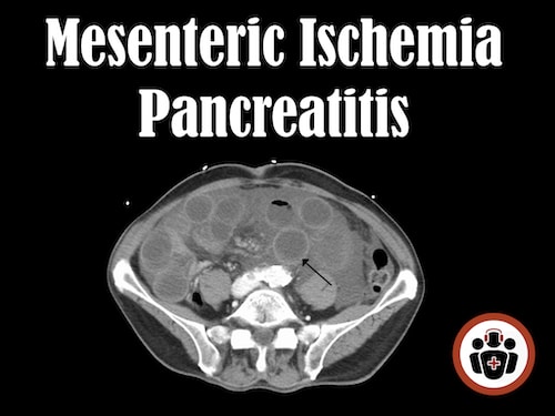
CT Abdomen for Mesenteric Ischemia
pooled sensitivity of 94% (95% CI = 90% to 97%) and specificity of 95% (95% CI = 93% to 97%). The positive likelihood ratio (+LR) for a positive CT = 17.5 (95% CI = 5.99 to 51.29), and the negative likelihood ratio (-LR) = 0.09 (95% CI = 0.05 to 0.17).
Neg LR = 0.09 – almost at 0.1 threshold of a rule out test!
CT – Speak with radiology regarding protocol:
- Venogram – if suspicion of venous thrombosis
- Angiogram – if suspicion of arterial emboli
- Triple phase (plain, venous and arterial phase CTs) – increased sensitivity for mesenteric ischemia; but, increased radiation exposure
Early CT Findings: non-specific findings, such as: bowel wall thickening, bowel dilation, mesenteric edema, ascites
*pitfall: to assume alternate Dx like infectious colitis when you get the non-specific early findings of mesenteric ischemia
Late CT Findings: pneumatosis, pneumoperitoneum, portal gas
ED Management of Mesenteric Ischemia
- Fluid resuscitation: can have massive 3rd space losses, +/- bleeding. Aggressive IV fluid resuscitation often required.
- Antibiotics: consider broad spectrum antibiotics if patient presents with a septic picture
- Anticoagulation (controversial): if embolic source, no urgent OR, and no bleeding, consider heparin
- Early surgical consult
- Vasopressors: try to avoid vasopressors in mesentric ischemia as they may worsen ischemia, but if required, choose pressors with least effect on splanchnic circulation (i.e. dobutamine/ milrinone). Avoid epinephrine, phenylephrine because of vasoconstrictive effects.
POST-ERCP ABOMINAL PAIN
- Pancreatitis: worsening abdominal pain, amylase 3x upper limit of normal, usually presents within 24h of ERCP
- Infection: i.e. ascending cholangitis
- Perforation: often retroperitoneal
- Bleeding
Rise in amylase should begin to washout within 3 days of ERCP, lipase may stay longer. Can be affected by CrCl.
PANCREATITIS
Presentation of Pancreatititis
Epigastric pain, can be RUQ or LUQ with radiation to back, relieved by sitting up (like pericarditis – can think of pancreatitis as ‘the pericarditis of the belly’)
Vomiting, +/- jaundice, abdo distension, ileus.
Pancreatitis Scoring Systems
clinical judgement has been shown to underestimate the severity of pancreatitis; hence scoring systems may be useful for prognosticiation
- APACHE II & Ranson Score- ICU setting, not applicable to ED
- CT severity index – based on degree of pancreatic necrosis seen on CT – may help prognosticate
- BISAP score (Bedside Index of Severity for Acute Pancreatitis): 1 pt for: BUN >5, GCS<15, 2 or more SIRS criteria, age >60, pleural effusion. Score 0 = 0% mortality, > 5 = 22% mortality – moderate utility in predicting who may need ICU monitoring but does not help decide who can be discharged or if better than clinical gestalt; performs as well as APACHE ll & Ranson but more applicable to ED and easier to calculate
For more on the BISAP score see Michelle Lin’s Academic Life in Emergency Medicine Blog – BISAP, EHMRG, ORT: 3 New Medical Scores You’ve Never Heard Of
Clues to sort out the Cause of Pancreatitis
- Alcohol pancreatitis: diffuse, gradual pain, AST > ALT (2:1 ratio)
- Gallstone pancreatitis: often RUQ pain, sudden onset, rise in ALT
Imaging Pancreatitis
- Ultrasound is indicated for first time diagnosis: helps determine if gallstones are the cause and if ERCP could be indicated.
- CT usually not helpful unless unsure of diagnosis, or suspect fulminant severe disease >48hrs since onset: CT is often normal within the first 48h; not test of choice to disgnose gallstones
Amylase & Lipase in Pancreatitis
- Amylase: sensitivity (about 80%) shorter t1/2 than lipase, therefore less reliable if presenting later in time course of disease.
- Lipase: sensitivity (about 90%) better sensitivity vs amylase for pancreatitis.
- Absolute number of lipase or amylase does not correlate to severity of disease.
- False elevation of amylase and lipase in renal failure.
for more on amylase vs lipase see Michelle Lin’s Blogpost at Academic Life in Emergency Medicine
Now Test Your Knowledge
![]() For more on mesenteric ischemia & pancreatitis on EM Cases:
For more on mesenteric ischemia & pancreatitis on EM Cases:
Best Case Ever 21 Abdominal Pain – Thinking Outside the Box
Dr. Helman, Dr. Steinhart and Dr. Dushenski have no conflicts of interest to declare.
References
- Acosta et al. (2012). J Emerg Med, 42(6): 635-41. www.ncbi.nlm.nih.gov/pubmed/?term=Acosta+et+al.+…
- Cudnik et al. (2013). Acad Emerg Med, 20(11): 1087-1100. www.ncbi.nlm.nih.gov/pubmed/?term=Cudnik+et+al.+…
- Papachristou et al. (2010). Am J Gastroenterology, 105(2): 435-41. www.ncbi.nlm.nih.gov/pubmed/?term=Papachristou+e…
- Sutton et al. (2009) Ann R Coll Surg Engl, 91(5): 381-4. www.ncbi.nlm.nih.gov/pubmed/?term=Sutton+et+al.+…
- Yadav et al. (2002). Am J Gastroenterology, 97(6): 1309-18. www.ncbi.nlm.nih.gov/pubmed/?term=Yadav+et+al.+(…
Now test your knowledge with a quiz.


Definitely this helped me a lot! thanks and keep it up!