
In this ECG Cases blog we assess 10 patients who presented with potential atrial fibrillation or atrial flutter. What is the diagnosis and best management?
Written by Jesse McLaren; peer Reviewed and edited by Anton Helman, February 2022
10 patients presented with possible atrial fibrillation/flutter. What’s the diagnosis and the management?
Case 1: 70yo, no prior atrial fibrillation, feeling weak for one day, normal vitals. Old then new ECG


Case 2: 70yo, history of atrial fibrillation taking anticoagulants, fall at home and found two days later. HR 150, BP 120/70 RR 20 oxygen saturation 96% temp 36.6

Case 3: 65yo, no history of atrial fibrillation, with 5 days shortness of breath on exertion and pleuritic chest pain. HR 115 BP 130/80 RR 20 oxygen saturation 95% temp 36.5

Case 4: 30yo two months postpartum with palpitations for 6 hours. HR 115 BP 110/80 RR 18 oxygen saturation 100% temp 36.4

Case 5: 80yo, history or CAD, with 2 hours chest pressure. HR 140 BP 120/75 RR 16 oxygen saturation 95% temp 36.4. Old then new ECG


Case 6: 70yo with two days on/off chest pain, suddenly worse over an hour. HR 120 BP 110/80 RR 20 oxygen saturation 96% temp 37 Old then new ECG


Case 7: 75yo, history of atrial fibrillation but not taking anticoagulants, with 10 hours of chest pain. HR 110, BP 120/80 RR 20, oxygen saturation 92%. Old then new ECG.


Case 8: 60yo history of diabetes, hypertension and atrial fibrillation, on beta-blocker and DOAC, with palpitations. HR 120, BP130 RR 18 oxygen saturation 95% temp 37.2

Case 9: 65yo history of CAD, CVA and paroxysmal atrial fibrillation, takes DOAC daily, with 7 hours of chest pressure. HR 120 BP 120/80. Old then new ECG


Case 10: 65yo history of hypertension and diabetes, with 4 hours palpitations. HR 150 BP130/80 R16 sat 99% T36.0

Approach to ECG interpretation of Atrial fibrillation
Atrial fibrillation/flutter is the most common dysrhythmia we treat in the emergency department, and there is a lot of room for quality improvement. As a recent study found, atrial fibrillation has high rates of overtreatment and undertreatment: a third of patients with atrial fibrillation secondary to an underlying medical condition received rate control rather than treatment of the underlying cause, while 40% of those with primary atrial fibrillation did not receive sufficient rate control to achieve a normal heart rate on discharge; and 10% with stroke risk factors were not discharge on anticoagulation.[1] This highlights the importance of standardization to improve the quality of care of patients with atrial fibrillation, as summarized in the CAEP AF best practices checklist.[2] As EM atrial fibrillation expert Dr. Ian Stiell has demonstrated, implementation of this checklist has been associated with increased rhythm control and reduced length of stay without any increase in adverse events.[3]
AFIB mnemonic
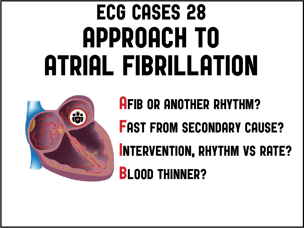
The approach to atrial fibrillation involves a series of questions summarized by the mnemonic AFIB:
- AF or another rhythm?
- Fast from a secondary cause?
- Intervention for primary AF: rhythm or rate control?
- Blood thinner?
Question 1: is it atrial fibrillation?
Before treating atrial fibrillation, the first step is ECG rhythm interpretation: is it actually atrial fibrillation or is it another dysrhythmia or simply artifact? Automated ECG interpretation is inaccurate in general, and has a high rate of error when it comes to atrial fibrillation. This includes overdiagnosis (applying the label of AF to what is actually sinus dysrhythmia, sinus tachycardia with premature atrial contraction, and/or baseline artifact) and underdiagnosis (applying the label of sinus tachycardia or SVT to what is actually AF, especially flutter).[4]
Question 2: if it is atrial fibrillation and there is rapid ventricular response, is it fast from a secondary cause?
This is important for diagnosis, management and prognosis. As EM atrial fibrillation expert Dr. Clare Atzema has found, patients with atrial fibrillation and an alternate primary diagnosis have three times the mortality rate compared with primary atrial fibrillation.[5] This is where the CAEP atrial fibrillation checklist starts: aggressively treat secondary causes (eg sepsis, GI bleed, PE, CHF, ACS, alcohol withdrawal), and avoid aggressive rate control in these patients.
One conundrum often arises with atrial fibrillation and ACS. As Dr. Atzema notes, 20% of patients with rapid AF experience chest pain, “but in the majority this is demand-related and not due to a rupture of an atherosclerotic plaque…The ECG findings before and subsequent to rate or rhythm control should be incorporated into the assessment.”[6] This also relates to the pattern of ischemic changes. Diffuse ST depression with reciprocal ST elevation in aVR is a non-specific sign of subendocardial ischemia that can be secondary to rapid atrial fibrillation—from secondary causes (like GI bleed or sepsis) or primary atrial fibrillation—and can be reassessed after treatment. Whereas rate controlled atrial fibrillation with regional and reciprocal ST/T changes is more concerning for acute coronary occlusion. While this is usually from plaque rupture, patients with atrial fibrillation are also at risk for coronary embolism, especially in those who are not anticoagulated.[7] Automated interpretation is also unreliable in this step, not only because of the limits of STEMI criteria to identify Occlusion MI, but also because they can apply the label “STEMI” to what is simply atrial flutter with fluctuating baseline.
Question 3: if it is atrial fibrillation with a rapid ventricular response without a secondary cause, what is the intervention?
Patients who are unstable as a result of acute primary atrial fibrillation require cardioversion, but as the checklist notes this is rare except for cases of atrial fibrillation with pre-excitation like WPW, and should prompt consideration of secondary causes. Stable patients with primary atrial fibrillation have the option of rhythm or rate control based on current anticoagulation, risk factors, and duration of symptoms. According to the checklist, it is safe to cardiovert if:
- Adequate anticoagulation for 3 weeks, or
- No history of TIA/CVA, no valvular heart disease and
- Onset <12 hours, or
- Onset 12-48 hours and <2 CHADS-65 (CHF, hypertension, age ≥ 65, diabetes), or
- No thrombus on transesophageal echo
As the RAFF2 trial found, both drug-shock (procainamide 15mg/kg followed by cardioversion if necessary for the 50% that don’t cardiovert with procainamide) and shock-only were highly effective at rhythm control.[8] Rate control for patients for whom cardioversion is unsafe includes calcium channel blockers or beta-blockers, and digoxin if hypotension or acute heart failure.
Question 4: does the patient need an anticoagulant started in the ED?
According to the checklist, anticoagulation is indicated (DOACs preferred over warfarin) if there are any CHADS-65 risk factors. EM thrombosis expert Dr. Kerstin de Wit has summarized contraindications to anticoagulation in the “McMaster checklist,” which is included in the CAEP best practices document. This includes absolute contraindications such as active serious bleeding, relative contraindications such as liver cirrhosis and renal failure (note that DOAC dose adjustments for patients with stable chronic kidney disease is often acceptable – speak with your pharmacist/hematologist).
Back to the cases
Case 1: artifact, no treatment needed
- Heart rate/rhythm: artifact, revealed by inconsistent ‘flutter’ waves in limb leads, which don’t correlate with precordial leads
- Electrical conduction: normal
- Axis: normal
- R-wave: old early R wave transition
- Tall/small voltages: normal voltages
- ST/T: old anterior ST depression
Impression: baseline artifact. Repeat ECG less artifact with visible P waves. Had negative workup

Case 2: atrial fibrillation with rapid ventricular response secondary to hypovolemia, treated with fluid resuscitation
- H: atrial flutter with rapid ventricular response and variable block
- E: LBBB
- A: normal axis
- R: delayed R wave progression from LBBB
- T: normal voltage
- S: appropriate discordant ST changes
Impression: atrial fibrillation secondary to hypovolemia. POCUS revealed hyperdynamic left ventricle and no free fluid in the abdomen to suggest bleeding from a fall while on anticoagulation. After fluid resuscitation the heart rate slowed down and rate-related LBBB resolved. Septic workup was negative, and the patient was admitted for dehydration.

Case 3: Atrial fibrillation secondary to pulmonary embolism, treated with anticoagulation
- H: AF with rapid ventricular response
- E: otherwise normal conduction
- A: physiologic left axis
- R: late R wave progression
- T: normal voltage
- S: no ST/T changes
Impression: atrial fibrillation with pleuritic chest pain and shortness of breath. D-dimer positive and CT chest revealed pulmonary embolism. Started on low molecular weight heparin and admitted. Remained in atrial fibrillation, and discharged on beta-blocker and DOAC.
Case 4: atrial fibrillation secondary to postpartum thyroiditis, treated with propranolol
- H: AF with rapid ventricular response
- E: otherwise normal conduction
- A: normal axis
- R: normal R wave
- T: low voltage
- S: no ST/T changes
Impression: atrial fibrillation postpartum. Had negative D-dimer, undetectable TSH was admitted with hyperthyroidism. Had normal echo, high T3/T4, and diagnosed with postpartum thyroiditis. Started on propranolol for rate control, no anticoagulation due to lack of CHADS-65 risk factors.
Case 5: primary atrial fibrillation with rapid ventricular response and rate-related subendocardial ischemia, unnecessary cath lab activation
- H: atrial fibrillation with rapid ventricular response
- E: otherwise normal conduction
- A: physiologic left axis
- R: old early R wave progression
- T: LVH
- S: diffuse STD with reciprocal STE-aVR
Impression: atrial fibrillation with rapid ventricular response and rate-related subendocardial ischemia. Cath lab was activated prior to rate control. No obstructive lesions, troponin negative, and ST changes resolved after spontaneous conversion.

Case 6: atrial fibrillation with inferoposterior MI, treated with reperfusion
- H: atrial fibrillation with rapid ventricular response
- E: otherwise normal conduction
- A: normal axis
- R: old early R wave transition
- T: normal voltage
- S: anterior ST depression, inferior Q waves
Impression: atrial fibrillation with inferoposterior MI. This could be from Occlusion MI, or Non-Occlusive MI with demand ischemia from rapid atrial fibrillation. Cath lab activated: 99% circumflex occlusion, first trop I was 3,000 (normal <26) and peak 43,000. Post-stent had spontaneous conversion, resolution of anterior ST depression, ongoing inferior Q waves. Discharged on DOAC in addition to antiplatelets.

Case 7: atrial fibrillation with first diagonal occlusion from coronary embolism, treated with reperfusion
- H: atrial fibrillation with mildly rapid ventricular response
- E: otherwise normal conduction
- A: normal axis
- R: delayed R wave progression
- T: normal voltage
- S: Q wave with ST elevation and T wave inversion (subacute occlusion) in I/aVL/V2 and reciprocal ST depression in III/aVF
Impression: first diagonal occlusion in patient with AF without anticoagulation. Cath lab activated: 100% first diagonal occlusion from coronary embolism. First troponin 25,000 and peak 48,000. Discharge ECG had resolution of ST changes and deeper reperfusion T wave inversion. Started on DOAC and antiplatelet.

Case 8: primary persistent atrial flutter, treated with rate control
- H: atrial flutter
- E: otherwise normal conduction
- A: right axis
- R: late R wave progression
- T: normal voltage
- S: no significant ST changes – flutter waves mislabeled as ST elevation
Impression: atrial flutter. Negative workup and rate controlled with beta-blocker

Case 9: primary paroxysmal atrial fibrillation, cardioverted with procainamide
- H: atrial fibrillation with rapid ventricular response
- E: old LAFB
- A: left axis from LAFB
- R: old delayed R wave progression, old Q in aVL/V2
- T: LVH
- S: no new ST/T changes
Impression: primary paroxysmal atrial fibrillation. History of CVA but on DOAC and has not missed any doses. Cardioverted with procainamide, negative workup and discharged.

Case 10: primary atrial flutter, electrically cardioverted
- H: atrial flutter with 2:1 conduction (negative flutter waves in lead II) – not sinus tachycardia
- E: otherwise normal conduction
- A: normal axis
- R: normal R wave progression
- T: normal voltage
- S: no ST/T changes
Impression: atrial flutter, no obvious secondary causes. Procainamide slowed down the ventricular response but did not cardiovert, so the patient received electrical cardioversion and was discharged on DOAC.


Early vs delayed cardioversion EM Quick Hit with Paul Dorion
Atrial fibrillation anticoagulation EM Quick Hit with Clare Atzema
Take home points on ECG approach to atrial fibrillation
- Atrial fibrillation? Computer interpretations have high rate of overdiagnosis (sinus arrhythmia, PAC, baseline artifact) and underdiagnosis (sinus tach, SVT)
- Fast from a secondary cause? Look for and treat underlying cause, eg. dehydration, sepsis, bleeding, PE
- Intervention? Rhythm or rate control based on stability, anti-coagulation, risk factors and duration
- Blood thinner? If CHADS-65 risk factors and no contraindications
References for ECG Cases 28: ECG approach to atrial fibrillation
- Wong BM, Green MS, Stiell IG. Rate Control Management of Atrial Fibrillation With Rapid Ventricular Response in the Emergency Department. Can J Cardiol 2020 Apr;36(4):509-517
- Stiell IG, de Wit K, Scheuermeyer FX, et al. 2021 CAEP Acute Atrial Fibrillation/Flutter Best Practices Checklist. CJEM 2021 Sep;23(5):604-610
- Stiell IG. RAFF-3 Trial: a stepped-wedge cluster randomized trial to improve care of acute atrial fibrillation and flutter in the emergency department. Can J Cardiol 2021 Jul 1;S0828-282X(21)00351-2
- Bae MH, Lee JH, Yang DH, et al. Erroneous computer electrocardiogram interpretation of atrial fibrillation and its clinical consequences. Clin Cardiol 2012 Jun;35(6):348-53
- Atzema CL, Lam K, Young C et al. Patients with atrial fibrillation and an alternative primary diagnosis in the emergency department: a description of their characteristics and outcomes. Acad Emerg Med 2013;20:193-199
- Atzema CL, Barrett TW. Managing atrial fibrillation. Ann Emerg Med 2015 May;65(5):532-9
- Shibata T, Kawakami S, Noguchi T, et al. Prevalence, clinical features, and prognosis of acute myocardial infarction attributable to coronary artery embolism. Circ 2015;132:241-250
- Stiell IG, Sililotti MCA, Taljaard M, et al. Electrical versus pharmacological cardioversion for emergency department patients with acute atrial fibrillation (RAFF2): a partial factorial randomised trial. Lancet 2020 Feb 1;395(10221):339-349


Excelent, a very proffessional approach
Very informative,logically and scientifically approached