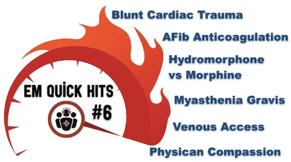
Topics in this EM Quick Hits podcast
Andrew Petrosoniak on diagnosis and risk stratification of blunt cardiac trauma (0:33)
Clare Atzema on latest guidelines for anticoagulation in atrial fibrillation (8:53)
Maria Ivankovic on hydromorphone vs morphine for acute pain (19:08)
Brit Long on clinical pearls in the diagnosis of myasthenia gravis (26:37)
Anand Swaminathan on venous access tips and tricks (30:11)
Bonus material from EM Cases Course June 2018 with Walter Himmel and Barbara Tatham on preventing burnout and physician compassion (podcast only).
Podcast production, editing and sound design by Anton Helman
Podcast content, written summary & blog post by Andrew Petrosoniak, Anand Swaminathan, Brit Long, edited by Anton Helman
Cite this podcast as: Helman, A. Swaminathan, A. Long, B. Petrosoniak, A. Atzema, C. Ivankovic M. EM Quick Hits 6 – Blunt Cardiac Trauma, Atrial Fibrillation Anticoagulation, Hydromorphone vs Morphine, Myasthenia Gravis, Venous Access. Emergency Medicine Cases. July, 2019. https://emergencymedicinecases.com/em-quick-hits-july-2019/. Accessed [date].
Diagnosis and Risk Stratification of Blunt Cardiac Trauma
- There is no gold standard definition of blunt cardiac injury. The clinician must use clinical judgement to decide based on clinical course and mechanism.
- If you suspect blunt cardiac injury, a negative ECG and troponin are sufficient to rule it out.
- A positive troponin in trauma predicts worse outcomes but it does not necessarily indicate blunt cardiac injury. There is little evidence to guide how to manage patients with elevated troponins following a trauma and the conservative approach is short admission for echo and telemetry.
- No need to worry about isolated sternal fracture, these really are predictably benign.
- Troponin is not a required test for all trauma patients unless there’s a feature that is concerning for blunt cardiac injury. Based on expert opinion and literature review, these include:
-
- Dysrhythmias
- Abnormal echocardiogram
- Unexplained hypotension
- Or CT evidence of significant mediastinal or cardiac injury
- Unexplained and persistent tachycardia
- Joseph B et al. Identifying the broken heart: predictors of mortality and morbidity in suspected blunt cardiac injury. Am J Surg 2016, 211, 982-88.
- Kalbitz M et al. The role of troponin in blunt cardiac injury after multiple trauma in humans. World J Surg 2017 Jan;41(1): 162-169.
- Odell DD et al. Sternal fracture: isolated lesion versus polytrauma from associated extrasternal injuries – analysis of 1867 cases. J Trauma Acute Care Surg 2013 Sep; 75(3):448-52.
Canadian Guideline Recommendations on Anticoagulation in Atrial Fibrillation
- Rather than considering cardioversion of atrial fibrillation within 48 hours of onset of atrial fibrillation without anticoagulation for all patients, guidelines recommend safe cardioversion without anticoagulation only for the patients with the lowest risk profile for stroke – those with a CHADS-2 <2. If CHADS-2 is ≥2 only cardiovert if onset of atrial fibrillation is within 12 hrs. The longer the duration of atrial fibrillation the higher the risk of stroke if cardioverted.
- The CCS Guidelines recommend anticoagulating all patients who are cardioverted regardless of stroke risk for a minimum of 4 weeks based on expert opinion only, while the CAEP checklist and AHA guidelines do not make this recommendation. While the risk of major bleeding is extraordinarily low for patients who are anticoagulated for only 4 weeks and the risk of stroke is also extraordinarily low for patients who are CHADS-65 negative or CHADS-2 <2, short term anticoagulation after cardioversion should be considered in low risk patients on an individual basis, incorporating shared decision making.
- All patients who are CHADS-65 positive or CHADS-2 ≥2 should be anticoagulated immediately for cardioversion or attempted cardioversion in the ED.
- Andrade JG, Verma A, Mitchell LB, et al. 2018 Focused Update of the Canadian Cardiovascular Society Guidelines for the Management of Atrial Fibrillation. Can J Cardiol. 2018;34(11):1371-1392.
- Stiell, I et al. CAEP Acute Atrial Fibrillation/Flutter Best Practices Checklist. CJEM 2018;20(3):334-342.
- Airaksinen KE, Grönberg T, Nuotio I, et al. Thromboembolic complications after cardioversion of acute atrial fibrillation: the FinCV (Finnish CardioVersion) study. J Am Coll Cardiol. 2013;62(13):1187-92.
- Stiell, Ian G. et al. Safe Cardioversion for Patients with Acute-Onset Atrial Fibrillation and Flutter: Practical Concerns and Considerations. Canadian Journal of Cardiology. Published online June 13, 2019.
Hydromorphone vs Morphine for Acute Pain
- Both ED use and prescriptions for hydromorphone have increased in the past decade in North America, while morphine prescriptions have remained stable.
- Hydromorphone has a faster onset with greater euphoric effects and potential for abuse compared to morphine, as well as higher street value, but equal analgesic effect with correct dosing.
- There is evidence to suggest that hydromorphone may be associated with a higher rate of adverse events compared to morphine including desaturations and inadvertent overdoses.
- Emergency physicians are more likely to under-dose morphine and to over-dose hydromorphone.
- The equal analgesic dose of hydromorphone 1mg po = morphine 5 mg po and hydromorphone 1mg IV = morphine 7-11mg.
- There is no evidence to support the notion that hydromorphone causes less pruritus, nausea or constipation compared to morphine.
- Patients with renal insufficiency are at risk for adverse events with both hydromorphone and morphine. Oversedation and respiratory depression are the main adverse events with morphine and delirium, agitation, hallucinations, myoclonus and seizures are the adverse events with hydromorphone. Nonetheless, some experts believe that hydromorphone is the opioid analgesic of choice in patients with renal insufficiency.
-
Narcotics Monitoring System, MOHLTC http://opioidprescribing.hqontario.ca
-
Gulur P, Koury K, Arnstein P, et al., Morphine versus Hydromorphone: Does Choice of Opioid Influence Outcomes? Pain Research and Treatment, vol. 2015, Article ID 482081, 6 pages, 2015.
-
Walsh SL, Nuzzo PA, Lofwall MR, Holtman JR Jr. The relative abuse liability of oral oxycodone, hydrocodone and hydromorphone assessed in prescription opioid abusers. Drug Alcohol Depend. 2008;98(3):191–202.
-
Beaudoin F.L., Merchant R.C., Janicki A. Preventing iatrogenic overdose: a review of in-emergency department opioid-related adverse drug events and medication errors. Ann Emerg Med. 2015;65(4):423–431.
-
O’Connor AB, Rao A. Why do emergency providers choose one opioid over another? A prospective cohort analysis. J Opioid Manag. 2012 Nov-Dec;8(6):403-13.
-
Mazer-Amirshahi, M., S. Motov, and L.S. Nelson, Hydromorphone use for acute pain: Misconceptions, controversies, and risks. J Opioid Manag, 2018. 14(1): p. 61-71.
-
Murray A et al. Hydromorphone. Journal of Pain and Symptom Management, 2005. 29(5) p57 – 66.
-
Smith, M.T., Neuroexcitatory effects of morphine and hydromorphone: evidence implicating the 3-glucuronide metabolites. Clin Exp Pharmacol Physiol, 2000. 27(7): p. 524-8.
Clinical Pearls in the Diagnosis of Myasthenia Gravis
- Myasthenia Gravis presents with abnormal extraocular muscle function, ptosis, and fatigable proximal muscle strength.
- Sensation, reflexes (including pupillary reflexes) should be normal. If they are abnormal consider Guillain Barre Syndrome, botulism and spinal cord disease.
- The ice pack test is your go-to test for suspected myasthenia in the ED. Simply place the ice pack over the patient’s affected closed eye for 2 minutes, remove and observe for improvement in ptosis. The ice pack test has a sensitivity = 90% and specificity = 80% for the diagnosis of Myasthenia Gravis.
- Roper J, Fleming ME, Long B, Koyfman A. Myasthenia gravis and crisis: evaluation and management in the emergency department. J Emerg Med. 2017 Dec;53(6):843-853.
Venous Access Pearls
- Obtaining access is a critical step in resuscitation and it’s important that resuscitationists have multiple approaches to accomplishing the task.
- Peripherals are powerful. Short and fat = higher flow rates. 18G peripheral IV >>> CVL in terms of flow.
- Go IO early: “1, 2, IO”, meaning 2 shots at a peripheral line and then move to IO placement.
- Humeral IOs should be our go to location and the key is to keep the arm in internal rotation to prevent dislodgement.
-
EMRAP HD: EZ IO Placement
Bonus material from EM Cases Course June 2018 with Walter Himmel and Barbara Tatham on preventing burnout and physician compassion (podcast only).
People don’t remember what you say. People don’t remember what you do. They remember how you make them feel.
None of the authors have any conflicts of interest to declare


Hy, great teaching as usual from EM cases. You don’t imagine what a huge impact your work has on the way many doctors all over the world practice. Thank you to the whole EM team!
I was deeply , personally touched by Barbara’s words in the end of this podcast.
Because I feel it is unfair, sad, and because it can happen to anyone of us, anytime…because she articulated so well the meaning of the relationship patient doctor.
Barbara thank you for your courage, thank you for sharing your story.Your message is very powerful and it will stay with me, hopefully helping me to better care for patients. I do know from experience ,it is not the duration of a life that matters , but rather the amount of light that life brings into the world. Thank you for your light Barbara.
As scientists we like to find out the “ why” behind things . Unfortunately in real life, we find ourselves without finding an answer. We just have to surrender and accept it. Than we can finally find peace.
Hugs Barbara
A Romanian doctor working in France, a mum, a friend from far