
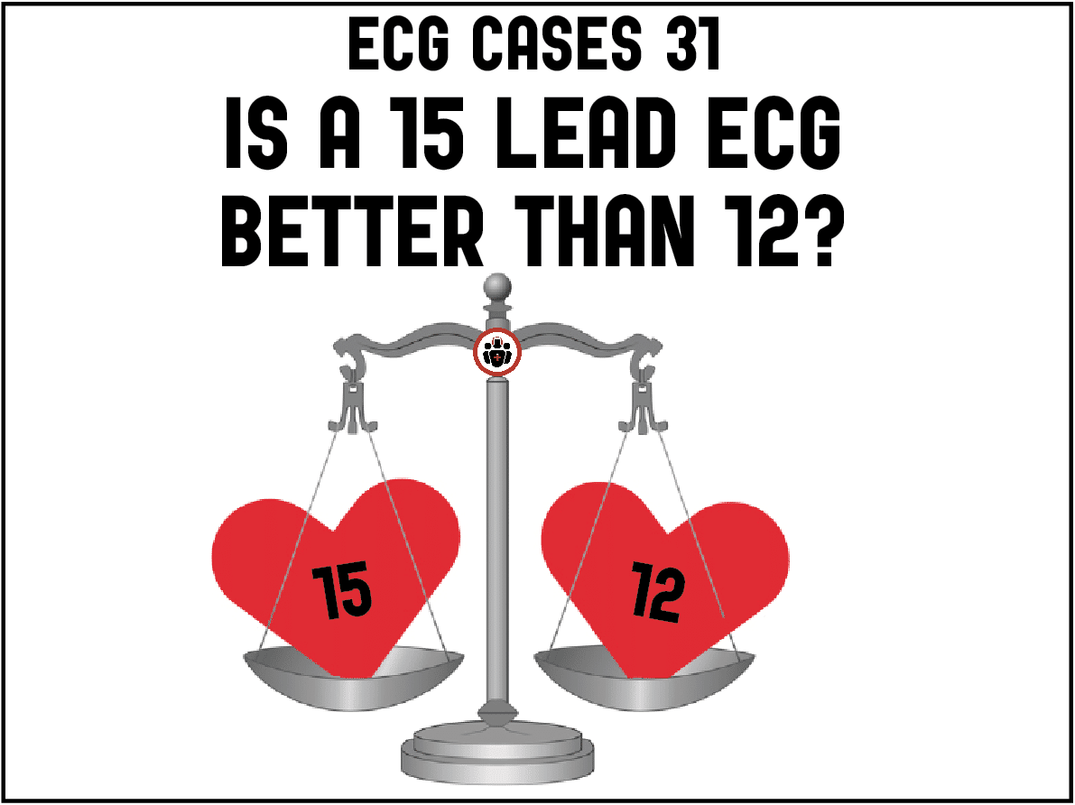
In this ECG Cases blog we review 8 patients with potentially ischemic symptoms. Do you need a 15-lead ECG? How will a 15-lead change management?
Written by Jesse McLaren; Peer Reviewed and edited by Anton Helman. May 2022
8 patients presented with potentially ischemic symptoms. Do you need a 15-lead ECG, and if so, how would it change management? How can we best diagnose posterior MI and RVMI on ECG?

Case 2: 65 year old with 24 hours on/off chest pain now constant, with dizziness. R20 sat 95% HR 60 BP 60/40

Case 3: 60yo with 4 hours of chest pain, nausea and syncope. R14 sat 100% HR 60 BP 90/60

Case 4: 50yo with one hour of chest pain radiating to the arms, R16 sat 99% HR 60 BP 150/90

Case 5: 65yo prior CABG with sudden chest pain and diaphoresis, normal vitals.

Case 6: 80 year old with VF arrest, return of spontaneous circulation with HR 100 and BP 100

Case 7: 65 year old with 8 hours of shoulder pain, normal vitals. Old then new ECG


Case 8: 60yo with exertional chest pain now constant, normal vitals. Old then new ECG:

12 vs 15 lead ECG: Diagnosing posterior MI and RVMI
The precordial ECG leads V1-V6 wrap around from the centre to the left side of the chest. But how can we diagnose infarctions involving the right ventricle or the posterior left ventricle? The 15-lead is supposed to provide a solution: the right sided lead V4R is placed on the right anterior chest (opposite the left chest lead V4) to look for right ventricular infarction, and the posterior leads V8-9 are added on the left midscapular and paraspinal to look for posterior MI. But routinely getting 15-lead ECGs on emergency department patients with chest pain does not improve diagnosis.[1] So what’s the role of the 15-lead ECG?
Before ordering any test, we need to ask: what clinical question are we trying to answer, and how will the test results change management. So before getting a 15-lead ECG, we need to know what question we are trying to answer, what information does the 12-lead already provide, and what are the added benefits or limitations of the additional leads.
Question 1: is there posterior Occlusion MI, and do you need V8-9?
The posterior (or basolateral) wall of the left ventricle is perfused by the right coronary or left circumflex artery. So posterior MI can be associated with inferior or lateral MI, or can be isolated posterior MI (from branches of the circumflex artery). The idea that we need posterior leads to identify posterior MI is based on three claims rooted in the STEMI paradigm. As a 1993 article explaining 15 lead ECGs summarized: 1) “ST-segment elevation…is the most specific marker of acute infarction, 2) “the impossibility of distinguishing anterior ‘subendocardial’ from posterior ‘transmural’ myocardial infarction; and 3) “leads V8 and V9 are superior to the diagnosis of posterior myocardial infarction to the reciprocal findings in leads V1 to V3.”[2].
But advances in ECG interpretation based on angiographic outcomes have led to a paradigm shift: ST elevation is not necessary to diagnose Occlusion MI (OMI), causes of ST depression can be differentiated, and the 12-lead is highly accurate for posterior MI. As a new study found, primary ischemic ST depression maximal in V1-V4 is 97% specific for posterior OMI, regardless of whether there are accompanying tall R waves or upright T waves (traditionally associated with posterior MI). This is distinct from secondary STD in the anterior leads (eg RVH, RBBB), or ischemic STDmaxV4-6 (from subendocardial ischemia). Posterior OMI is often associated with other signs of OMI including subtle ST elevation and hyperacute T waves in the inferior and/or lateral walls. As Meyers et al. noted, “all patients with posterior OMI had either (1) at least 1mm STD max V1-4 or (2) other subtle findings of OMI in addition to any STD max V1-4 (even if <1mm)…On the basis of our findings, we believe STD max V1-4 to be highly accurate and sufficient without the need for routine posterior leads.”[3]
So, to identify posterior OMI simply look at the 12 lead for primary ischemic STD max V1-4, and associated inferior/lateral signs. Posterior leads may show ST elevation but this is not required if the 12-lead is already diagnostic, and the low voltage posterior leads can also be falsely negative and lead to delayed diagnosis.
Question 2: is there right ventricular MI, and do you need V4R?
The right ventricle has dual blood supply from the right and left coronary arteries. If the RCA is occluded proximally, and there is insufficient collateral circulation, this can lead to RV myocardial infarction (RVMI) with the risk of hypotension and preload dependence. So RVMI changes prognosis and management, from nitro to fluids
Step 1
Usually the RCA also perfuses the LV inferior wall, so the first step to identifying RMVI is to identify inferior OMI on the 12-lead—with inferior ST elevation and hyperacute T waves, and reciprocal ST depression in aVL. While many inferior OMI don’t meet STEMI criteria, nearly all will have reciprocal change in aVL. Most inferior OMI are from the RCA rather than the circumflex—especially if there is ST depression in lead I (away from left sided leads), ST elevation in III (right inferior lead)>II (left inferior), and if the ST elevation inferiorly is greater than the ST depression anteriorly [4]. It’s important to start with inferior OMI, because ST elevation on V4R can also be seen in nonspecific subendocardial ischemia causing ST elevation in aVR, or anterior STEMI causing ST elevation in V1[5]. Rarely RVMI can be isolated, with STE V1-V3 on the 12 lead mimicking LV septal MI.
Step 2
The second step is looking for RV infarct on the 12-lead. Lead V1 is the most rightward of precordial leads on the 12 lead, so ST elevation in V1 in the presence of inferior OMI identifies RVMI with a specificity of 84%. But sensitivity is 69% and drops to 35% if there’s ST depression in V2. As a study by Bischof et al explained, “when there is concurrent posterior MI, as manifested by concurrent ST depression in lead V2: such ST depression would attenuate the ST elevation in lead V1, ‘pulling it down’ and resulting in a false negative (absence of STE in V1 in the presence of RVMI).” [6]
Step 3
If there is inferior OMI without ST elevation in V1 on the 12-lead, a 15-lead ECG can improve accuracy—with STE-V4R identifying proximal RCA occlusion with a sensitivity of 96% and specificity of 82%. But sensitivity drops over time, or in the presence of concomitant posterior OMI for the same reasons that limit the 12 lead: “The right ventricular anterior wall and posterior wall are diagonally opposite to each other, and the latter is thicker, producing greater injury current. Therefore, right precordial ST-segment elevation caused by RVI is offset by reciprocal changes in posterior ST-segment elevation caused by PWI, thereby masking the electrocardiographic sign of RVI, i.e.,ST↑V4R.” [7]
So, to identify RVMI we need to first start with inferior OMI, then look for associated ST elevation in V1, and if it’s not present a 15 lead can increase sensitivity (but is limited by concomitant posterior MI).
Back to the cases
- Heart rate/rhythm: low atrial rhythm (P wave upright in I but inverted in II)
- Electrical conduction: normal conduction
- Axis: normal axis
- R-wave progression: normal R wave progression except for Q in V1-2
- Tall/small voltages: normal voltages
- ST/T: V1-2 ST elevation and hyperacute T waves, with reciprocal inferolateral ST depression
Impression: STEMI(+)OMI, which could be LV septum or isolated RV
15 lead: back to sinus rhythm, falsely negative V4R, posterior lead reciprocal ST depression

Cath lab activated: 100% occlusion of the conus artery (first branch of the RCA) which could not be reopened, then cardiac arrest.
Case 2: proximal RCA occlusion with RVMI on 12 lead
- H: junctional rhythm (with retrograde P waves after the QRS), borderline bradycardia
- E: normal QRS/QT
- A: normal
- R: normal
- T: normal voltages
- S: inferior ST elevation with reciprocal STD-aVL (inferior OMI), STE III>II and STD in I (RCA), plus ST elevation in V1 (RVMI)
Impression: inferior STEMI(+)OMI with RVMI, complicated by junctional rhythm and hypotension. No 15 lead done but would not have changed management. EMS treated with aspirin and fluid bolus and directly to cath lab: 100% proximal RCA occlusion. First trop I was 10,000 ng/L (normal <26 in males, <16 in females) and peak 50,000. Discharge ECG had resolution of ST changes, appearance of inferior Q waves and development of inferior reperfusion T wave inversion:

Case 3: proximal RCA occlusion with RVMI seen better on 15 lead
- H: borderline sinus tach with complete AV block with variable junctional escape rhythm
- E: complete AV block, narrow QRS, normal QT
- A: normal axis
- R: normal R wave progression
- T: normal voltages
- S: inferior STE reciprocal STD-aVL (inferior OMI), STE III>II and STD in I (RCA), no STE in V1 but STD in V2-3 (posterior OMI)
Impression: infero-posterior STEMI(-)OMI, ST depression in V2 from posterior OMI could counteract STE in V1 from RVMI.
15 lead had true positive STE in V4R but false negative posterior leads

Treated with dual antiplatelets/heparin and fluids, cath lab activated: 100% proximal RCA occlusion, required temporary pacing for bradycardia. First troponin I was 36 and peak 47,000. Discharge ECG had return of normal sinus rhythm, resolution of ST changes and development of inferior reperfusion T wave inversion:

Case 4: inferoposterior OMI from mid RCA occlusion, V4R true negative and V8-9 not required
- H: normal sinus rhythm with premature junctional beats
- E: normal conduction
- A: normal axis
- R: normal R wave progression
- T: normal voltages
- S: inferior STE reciprocal TWI in aVL (inferior OMI), STE III>II and STD in I (RCA), and STD in V2 (posterior)
Impression: inferoposterior STEMI(-)OMI, no STE in V1 but there’s STD in V2
15 lead showed no STE in V4R, minimal STE in V9

Treated with dual antiplatelets/heparin and nitro, cath lab activated: triple vessel disease with 100% mid RCA occlusion. First trop I was 17 and peak was 50,000. Discharge ECG had reperfusion T wave inversion inferiorly, posteriorly and in V4-5:

Case 5: diffuse STD with reciprocal STE-aVR and false positive STE-V4R
- H: normal sinus rhythm
- E: normal conduction
- A: normal axis
- R: normal R wave
- T: normal voltages
- S: diffuse ST depression with reciprocal STE in AVR/V1
Impression: nonspecific but high risk subendocardial ischemia, with presentation consistent with acute coronary syndrome.
15 lead was done showing mild STE in V4R because also elevated in aVR and V1

Cath lab: 99% occlusion of posterior interventricular branch from distal RCA occlusion, not proximal RCA. First trop 62 and peak 40,000. Discharge ECG had inferior Q waves and reperfusion T wave inversion:

Case 6: 80 year old with VF arrest, return of spontaneous circulation with HR 100 and BP 100
- H: normal sinus rhythm
- E: normal conduction
- A: normal axis
- R: normal R wave progression
- T: small voltages in limb leads
- S: significant primary STD maximal in V1-4
Impression: posterior STEMI(-)OMI diagnostic on the 12 lead.
15 lead not required but done anyways, showing small voltages with minimal STE V8-9

Cath lab activated: 100% circumflex occlusion. Follow up ECG had tall anterior T waves from posterior reperfusion T wave inversion:

Case 7: posterior STEMI(-)OMI diagnostic on 12 lead, delayed diagnosis/treatment because posterior leads falsely negative
- H: normal sinus rhythm
- E: normal conduction
- A: normal axis
- R: normal R wave progression
- T: normal voltages
- S: primary STD max V1-V4, with subtle signs of inferior OMI (new inferior Q waves, minimal STE in III with reciprocal STD in I/aVL), anterior STD > inferior STE
Impression: inferoposterior STEMI(-)OMI, diagnostic on 12 lead.
15 lead had no ST elevation in V4R (true negative) and none in posterior leads (false negative), while still being diagnostic on the anterior leads:

Because ECG did not meet STEMI criteria, the patient was treated with asa and nitro and had stat cardiology consult. Cath lab only activated after troponin returned at 6,000, which was 90 minutes after the first ECG. There was 100% circumflex occlusion, but the patient was diagnosed as “NSTEMI”. No repeat troponins done. Discharge ECG had resolution of ST changes, and Q waves with reperfusion T wave inversion in III/aVF and V6:

Case 8: subtle posterior OMI, more obvious on posterior leads:
- H: normal sinus rhythm
- E: normal conduction
- A: borderline right axis
- R: normal R wave progression
- T: normal voltages
- S: pseudonormalization of ST segment V2-3
Impression: subtle posterior (STEMI-)OMI.
15-lead done, which showed more obvious STE:

Cath lab activated: 100% circumflex occlusion, first trop in normal range and peak 50,000
Take home points for 15-lead ECG and diagnosing posterior MI and RVMI
- Posterior OMI can be diagnosed on the 12-lead (primary STD max V1-4 +/- inferior or lateral OMI) and V8-9 can be falsely negative.
- RVMI (proximal RCA occlusion) is associated with hypotension and nitro sensitivity, and can be diagnosed on the 12-lead if inferior OMI also has STE in V1.
- There are 3 steps to diagnose RVMI on ECG:Step 1: identify inferior OMI on the 12-leadStep 2: look for RV infarct on the 12-lead – ST elevation in V1 in the presence of inferior OMI identifies RVMI with a specificity of 84%.Step 3: If there is inferior OMI without ST elevation in V1 on the 12-lead, a 15-lead ECG can improve accuracy—with STE-V4R identifying proximal RCA occlusion with a sensitivity of 96% and specificity of 82%, but can be falsely negative over time or in the presence of posterior OMI.
References for ECG Cases 31: Is a 15 lead ECG better than 12? Diagnosing Posterior MI and RVMI
- Brady WJ, Hwang V, Sullivan R, et al. A comparison of 12- and 15-lead ECGs in ED chest pain patients: impact on diagnosis, therapy and disposition
- Zalenski RB, Cooke D, Rydman R, et al. Assessing the diagnostic value of an ECG containing leads V4R, V8, and V9: the 15-lead ECG. Ann Emerg Med 1993 May;22(5): 786-93
- Meyers HP, Bracey A, Lee D, et al. Ischemic ST-segment depression maximal in V1-V4 (versus V5-V6) of any amplitude is specific for Occlusion Myocardial Infarction (versus nonocclusive ischemia). J of Amer Heart Assoc 2021;10:e022866
- Fiol M, Cygankiewicz I, Carrillo A, et al. Value of electrocardiographic algorithm based on ‘ups and downs’ of ST in assessment of a culprit artery in evolving inferior wall acute myocardial infaraction. Am J Cardiol 2004
- Dehghani P, Zahedi A, Hassanzadeh M, et al. Significance of ST-segment elvation in V4R lead in patients with anterior myocardial infarction. Ann Noninvasive Electrocardiol 2021
- Bischof JE, Worrall CI, Smith SW. In inferior myocardial infarction, neither ST elevation in lead V1 nor ST depression in lead I are reliable findings for the diagnosis of right ventricular infarction. J of Electrocardiol 2018
- Kosuge M, Ishikawa T, Morita S, et al. Posterior wall involvement attenuates predictive value of ST-segment elevation in lead V4R for right ventricular involvement in inferior acute myocardial infarction. J Cardiol 2009


Excellent ????????