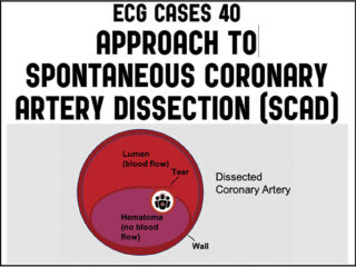
ECG Cases 40 – Approach to Spontaneous Coronary Artery Dissection (SCAD)

Dr. Jesse McLaren on when to consider Spontaneous Coronary Artery Dissection (SCAD), which patients are at risk for reocclusion, and the challenges of diagnosing SCAD in patients who have nonischemic ECGs despite silent occlusion, occlusions perfused by collaterals, or from non-occlusive MI on this ECG Cases...

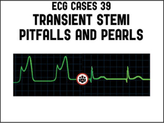
ECG Cases 39 – Transient STEMI Pitfalls and Pearls

In this ECG Cases blog we look at 9 patients with possible transient STEMI and discuss pitfalls and pearls in ECG interpretation and management...

ECG Cases 38 – ECG Interpretation in Cocaine Chest Pain

Dr. Jesse McLaren discusses some key aspects of cocaine chest pain ECG interpretation in this month's blog including: Patients with cocaine-associated chest pain require benzodiazepines +/- nitroglycerine for symptom relief, aspirin and ECG to look for signs of occlusion and reperfusion. In patients with chest pain + ST elevation, consider false positive STEMI including early repolarization, LVH and Brugada-pattern. In patients with cocaine chest pain who are STEMI negative, beware STEMI(-)OMI including subtle ST elevation, hyperacute T waves, reciprocal change, and refractory ischemia. For cocaine chest pain patients who's chest pain has resolved, look for reperfusion T wave inversion, as this may put them at risk for reocclusion.

ECG Cases 35 – ECG Approach to Takotsubo Syndrome

Takotsubo Syndrome is usually triggered by an emotional or physical stress leading to acute catecholaminergic myocardial stunning. The initial ST elevation phase of Takotsubo Syndrome mimics Occlusion MI, can not be distinguished by patient factors or POCUS findings, and requires immediate angiogram. The subsequent phase of Takotsubo Syndrome has T wave inversion in an apical distribution, which can mimic reperfusion, but often has very deep T wave inversions and a very long QT interval. Takotsubo Syndrome is a retrospective diagnosis of exclusion—with an angiogram ruling out occlusion, a ventriculogram showing apical ballooning, and a follow up echo showing recovery of LV function. Complications of Takotsubo Syndrome include LV failure, apical thrombus, and polymorphic VT from long QT. Jesse McLaren guides us through 10 ECGs to elucidate these important take home points...

ECG Cases 34 – ECG Interpretation in Aortic Dissection

Which patients with ECG evidence of coronary occlusion require a CT scan to rule out aortic dissection? What are the range of ECG findings in acute aortic dissection and how do they change management? Dr. Jesse McLaren guides us through 9 cases to answer these and other questions on ECG interpretation in aortic dissection...

ECG Cases 32 Prehospital ECG pearls and pitfalls

In this ECG Cases blog we review 8 cases of patients with prehospital ECGs and explore prehospital ECGs for diagnosing STEMI, Occlusion MI, false STEMI, code STEMI, dynamic ischemic changes, truncated voltages. Can you avoid the pitfalls and spot the pearls that help to make the diagnosis?

ECG Cases 31 Is a 15 lead ECG better than 12? Diagnosing Posterior MI and RVMI

Is 15 lead ECG better than 12 lead for diagnosing posterior MI or right ventricular infarction? When do you need a 15 lead ECG? Jesse McLaren guides us through 8 cases to highlight the steps and pitfalls in diagnosing posterior MI and RVMI in light of recent ECG literature...

ECG Cases 30 Beware Computer Interpretation Errors

Computer interpretation of the ECG has been called a double-edged sword: when correct, it increases physician accuracy, but when incorrect it increases errors. This is especially problematic in the emergency department, where computer accuracy drops as clinical significance increases—with common errors for arrhythmias and ischemia. Jesse McLaren guides us through 10 cases where the computer interpretation misguides us and how to avoid these pitfalls...

ECG Cases 29 Misdiagnosis from Lead Misplacement, Artifact and Lead Reversal

In this ECG Cases blog we review 10 cases of possible artifact, lead reversal and lead misplacement. Can you spot the abnormalities and avoid the misdiagnosis?...

ECG Cases 27 Pericarditis – Diagnosis of Exclusion

Jesse McLaren guides us through 9 cases and explains how pericarditis is a diagnosis of exclusion through 3 simple steps: 1. Exclude more serious causes of chest pain, eg wraparound LAD occlusion, inferior OMI 2. Exclude complications of pericarditis, eg myocarditis, large pericardial effusion 3. Exclude normal variant ST elevation presenting with benign chest pain on this month's ECG Cases blog...

Donate Subscribe
Go to Top